
上海细胞库
人源细胞系| 稳转细胞系| 基因敲除株| 基因点突变细胞株| 基因过表达细胞株| 重组细胞系| 猪的细胞系| 马细胞系| 兔的细胞系| 犬的细胞系| 山羊的细胞系| 鱼的细胞系| 猴的细胞系| 仓鼠的细胞系| 狗的细胞系| 牛的细胞| 大鼠细胞系| 小鼠细胞系| 其他细胞系|
人源细胞系| 稳转细胞系| 基因敲除株| 基因点突变细胞株| 基因过表达细胞株| 重组细胞系| 猪的细胞系| 马细胞系| 兔的细胞系| 犬的细胞系| 山羊的细胞系| 鱼的细胞系| 猴的细胞系| 仓鼠的细胞系| 狗的细胞系| 牛的细胞| 大鼠细胞系| 小鼠细胞系| 其他细胞系|

| 规格 | 价格 | 库存 |
|---|---|---|
| 50ul | ¥ 1580.00 | 10 |
| 100ul | ¥ 2500.00 | 10 |
| 产品编号 | bsm-52353R |
| 英文名称 | Rabbit?Anti-Hsp90 alpha ?antibody |
| 中文名称 | 热休克蛋白90α重组兔单抗 |
| 别????名 | HSP-90 alpha; HSP 86; HSP 86; Renal carcinoma antigen NY REN 38; Heat shock 86 kDa; Heat shock 90kDa protein 1 alpha; Heat shock protein 90kDa alpha (cytosolic) class A member 1; heat shock protein 90kDa alpha (cytosolic), class A member 2; Heat shock protein HSP 90-alpha; HS90A_HUMAN; HSP 86; HSP86; Hsp89; HSP89A; HSP90A; HSP90AA1; HSP90ALPHA; HSP90N; HSPC1; HSPCA; HSPCAL1; HSPCAL3; HSPCAL4; HSPN; LAP2; Renal carcinoma antigen NY-REN-38.?? |
| 研究领域 | 肿瘤??信号转导??转录调节因子?? |
| 抗体来源 | Rabbit |
| 克隆类型 | Recombinant |
| 交叉反应 | (predicted: Human,Mouse,Rat) |
| 产品应用 | WB=1:500-1000, IHC-P=1:100-500, ICC=1:50-200, IF=1:50-200 not yet tested in other applications. optimal dilutions/concentrations should be determined by the end user. |
| 理论分子量 | 85kDa |
| 细胞定位 | 细胞浆?细胞膜? |
| 性????状 | Liquid |
| 浓????度 | 1mg/ml |
| 免?疫?原 | KLH conjugated synthetic peptide derived from human Hsp90 alpha ? |
| 亚????型 | IgG |
| 纯化方法 | affinity purified by Protein A |
| 缓?冲?液 | 0.01M TBS(pH7.4) with 1% BSA, 0.03% Proclin300 and 50% Glycerol. |
| 保存条件 | Shipped at 4℃. Store at -20 °C for one year. Avoid repeated freeze/thaw cycles. |
| 注意事项 | This product as supplied is intended for research use only, not for use in human, therapeutic or diagnostic applications. |
| PubMed | PubMed |
| 产品介绍 | Hsp90 (heat shock protein 90) is a molecular chaperone and is one of the most abundant proteins in unstressed cells. It is an ubiquitous molecular chaperone found in eubacteria and all branches of eukarya, but it is apparently absent in archaea. Whereas cytoplasmic Hsp90 is essential for viability under all conditions in eukaryotes, the bacterial homologue HtpG is dispensable under non-heat stress conditions. Function: Molecular chaperone that promotes the maturation, structural maintenance and proper regulation of specific target proteins involved for instance in cell cycle control and signal transduction. Undergoes a functional cycle that is linked to its ATPase activity. This cycle probably induces conformational changes in the client proteins, thereby causing their activation. Interacts dynamically with various co-chaperones that modulate its substrate recognition, ATPase cycle and chaperone function. Subunit: Homodimer. Interacts with AHSA1, FNIP1, HSF1, SMYD3 and TOM34. Interacts with TERT; the interaction, together with PTGES3, is required for correct assembly and stabilization of the TERT holoenzyme complex. Interacts with CHORDC1 and DNAJC7. Interacts with STUB1 and UBE2N; may couple the chaperone and ubiquitination systems. Subcellular Location: Cytoplasm. Melanosome. Note=Identified by mass spectrometry in melanosome fractions from stage I to stage IV. Post-translational modifications: ISGylated. S-nitrosylated; negatively regulates the ATPase activity and the activation of eNOS by HSP90AA1. Similarity: Belongs to the heat shock protein 90 family. SWISS: P07900 Gene ID: 3320 Database links: Entrez Gene: 3320?Human Entrez Gene: 15519?Mouse Omim: 140571?Human SwissProt: P07900?Human SwissProt: P07901?Mouse Unigene: 525600?Human Unigene: 700831?Human Unigene: 1843?Mouse Unigene: 341186?Mouse Unigene: 119867?Rat 热休克蛋白-90α主要在类肌细胞、间质细胞、支持细胞核及生精细胞的胞浆表达;HSP-90 alpha与肿瘤的关系密切,在许多肿瘤细胞中HSP90α表达升高. |
| 产品图片 | 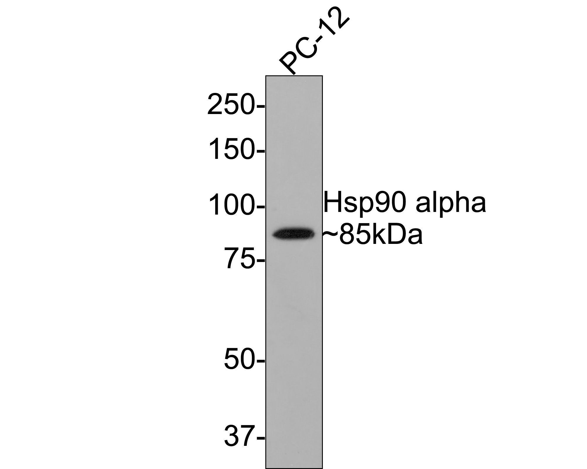 Western blot analysis of Hsp90 alpha on PC-12 cell lysates with Rabbit anti-Hsp90 alpha antibody (bsm-52353R) at 1/500 dilution. Lysates/proteins at 10 ?g/Lane. Predicted band size: 85 kDa Exposure time: 2 minutes; 8% SDS-PAGE gel. Proteins were transferred to a PVDF membrane and blocked with 5% NFDM/TBST for 1 hour at room temperature. The primary antibody (bsm-52353R) at 1/500 dilution was used in 5% NFDM/TBST at room temperature for 2 hours. Goat Anti-Rabbit IgG - HRP Secondary Antibody (HA1001) at 1:300,000 dilution was used for 1 hour at room temperature. 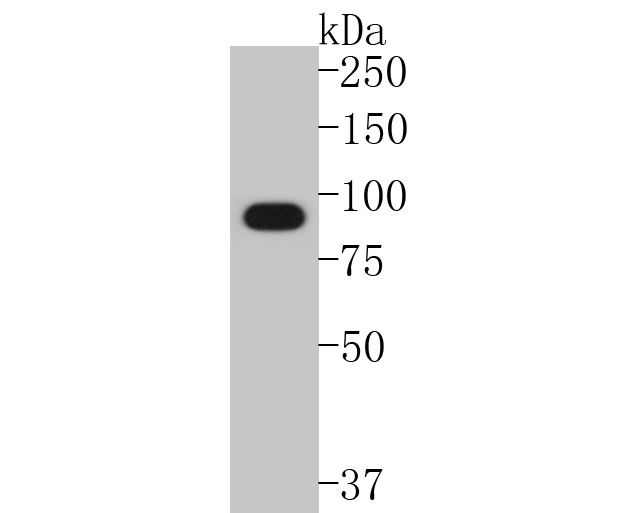 Western blot analysis of Hsp90 alpha on COS-1 cell lysates. Proteins were transferred to a PVDF membrane and blocked with 5% BSA in PBS for 1 hour at room temperature. The primary antibody (bsm-52353R, 1/500) was used in 5% BSA at room temperature for 2 hours. Goat Anti-Rabbit IgG - HRP Secondary Antibody (HA1001) at 1:5,000 dilution was used for 1 hour at room temperature. 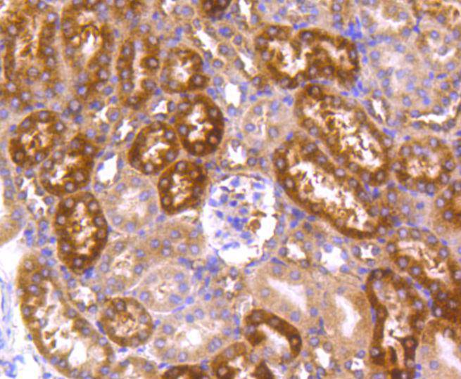 Immunohistochemical analysis of paraffin-embedded mouse kidney tissue using anti-Hsp90 alpha antibody. The section was pre-treated using heat mediated antigen retrieval with Tris-EDTA buffer (pH 8.0-8.4) for 20 minutes.The tissues were blocked in 5% BSA for 30 minutes at room temperature, washed with ddH2O and PBS, and then probed with the primary antibody (bsm-52353R, 1/50) for 30 minutes at room temperature. The detection was performed using an HRP conjugated compact polymer system. DAB was used as the chromogen. Tissues were counterstained with hematoxylin and mounted with DPX. 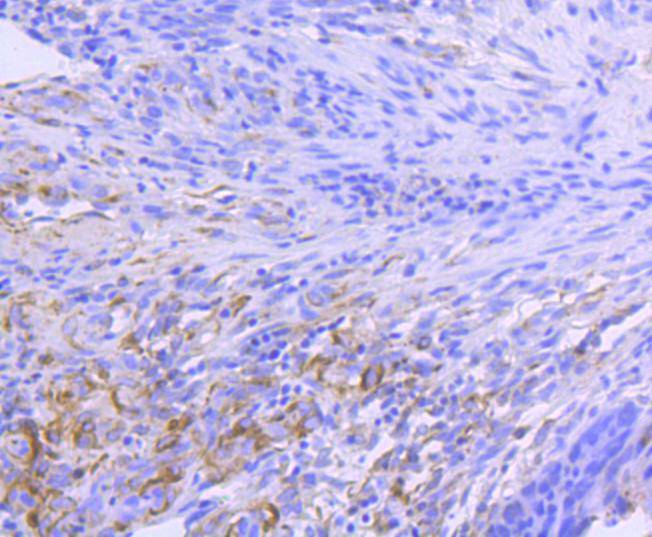 Immunohistochemical analysis of paraffin-embedded human colon carcinoma tissue using anti-Hsp90 alpha antibody. The section was pre-treated using heat mediated antigen retrieval with Tris-EDTA buffer (pH 8.0-8.4) for 20 minutes.The tissues were blocked in 5% BSA for 30 minutes at room temperature, washed with ddH2O and PBS, and then probed with the primary antibody (bsm-52353R, 1/50) for 30 minutes at room temperature. The detection was performed using an HRP conjugated compact polymer system. DAB was used as the chromogen. Tissues were counterstained with hematoxylin and mounted with DPX. 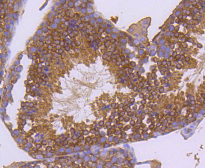 Immunohistochemical analysis of paraffin-embedded mouse testis tissue using anti-Hsp90 alpha antibody. The section was pre-treated using heat mediated antigen retrieval with Tris-EDTA buffer (pH 8.0-8.4) for 20 minutes.The tissues were blocked in 5% BSA for 30 minutes at room temperature, washed with ddH2O and PBS, and then probed with the primary antibody (bsm-52353R, 1/50) for 30 minutes at room temperature. The detection was performed using an HRP conjugated compact polymer system. DAB was used as the chromogen. Tissues were counterstained with hematoxylin and mounted with DPX. 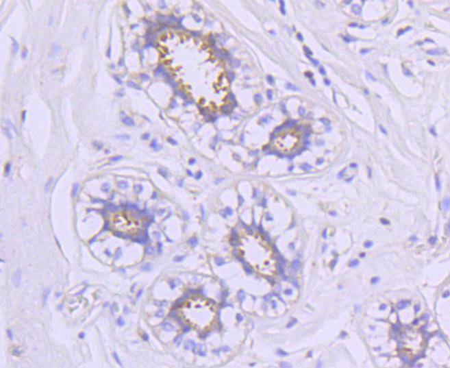 Immunohistochemical analysis of paraffin-embedded human breast tissue using anti-Hsp90 alpha antibody. The section was pre-treated using heat mediated antigen retrieval with Tris-EDTA buffer (pH 8.0-8.4) for 20 minutes.The tissues were blocked in 5% BSA for 30 minutes at room temperature, washed with ddH2O and PBS, and then probed with the primary antibody (bsm-52353R, 1/50) for 30 minutes at room temperature. The detection was performed using an HRP conjugated compact polymer system. DAB was used as the chromogen. Tissues were counterstained with hematoxylin and mounted with DPX. 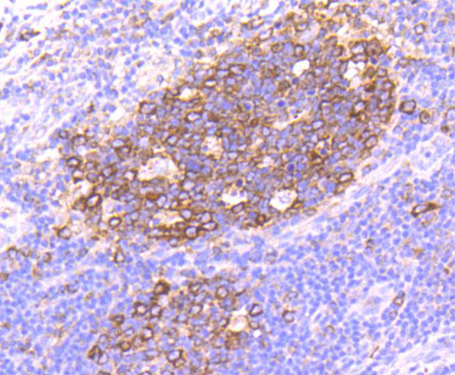 Immunohistochemical analysis of paraffin-embedded human tonsil tissue using anti-Hsp90 alpha antibody. The section was pre-treated using heat mediated antigen retrieval with Tris-EDTA buffer (pH 8.0-8.4) for 20 minutes.The tissues were blocked in 5% BSA for 30 minutes at room temperature, washed with ddH2O and PBS, and then probed with the primary antibody (bsm-52353R, 1/50) for 30 minutes at room temperature. The detection was performed using an HRP conjugated compact polymer system. DAB was used as the chromogen. Tissues were counterstained with hematoxylin and mounted with DPX. 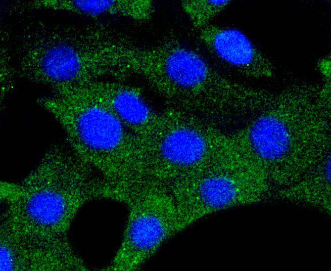 ICC staining of Hsp90 alpha in NIH/3T3 cells (green). Formalin fixed cells were permeabilized with 0.1% Triton X-100 in TBS for 10 minutes at room temperature and blocked with 1% Blocker BSA for 15 minutes at room temperature. Cells were probed with the primary antibody (bsm-52353R, 1/50) for 1 hour at room temperature, washed with PBS. Alexa Fluor?488 Goat anti-Rabbit IgG was used as the secondary antibody at 1/1,000 dilution. The nuclear counter stain is DAPI (blue). 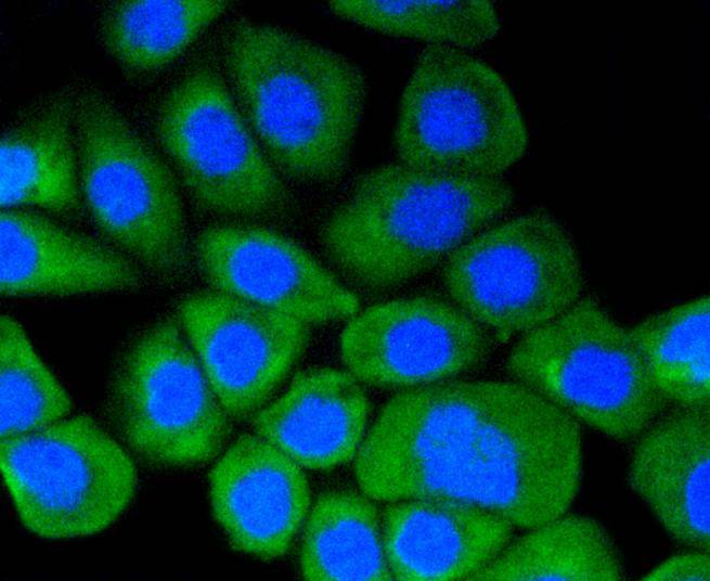 ICC staining of Hsp90 alpha in AGS cells (green). Formalin fixed cells were permeabilized with 0.1% Triton X-100 in TBS for 10 minutes at room temperature and blocked with 1% Blocker BSA for 15 minutes at room temperature. Cells were probed with the primary antibody (bsm-52353R, 1/50) for 1 hour at room temperature, washed with PBS. Alexa Fluor?488 Goat anti-Rabbit IgG was used as the secondary antibody at 1/1,000 dilution. The nuclear counter stain is DAPI (blue). 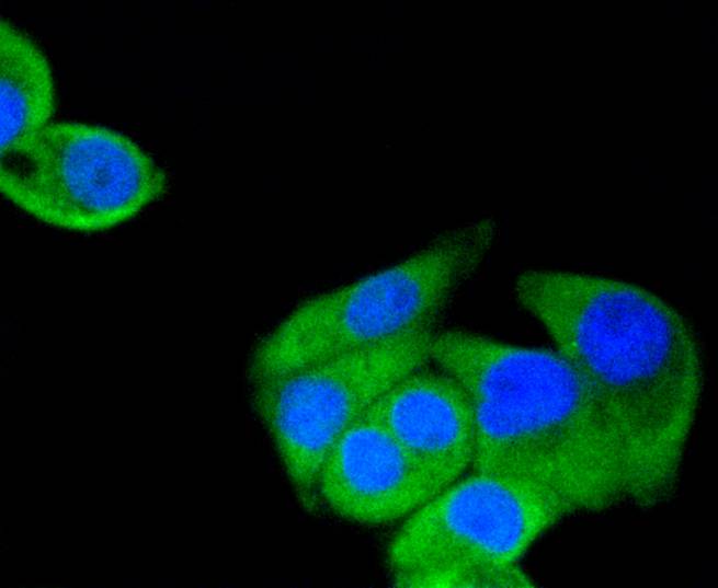 ICC staining of Hsp90 alpha in Hela cells (green). Formalin fixed cells were permeabilized with 0.1% Triton X-100 in TBS for 10 minutes at room temperature and blocked with 1% Blocker BSA for 15 minutes at room temperature. Cells were probed with the primary antibody (bsm-52353R, 1/50) for 1 hour at room temperature, washed with PBS. Alexa Fluor?488 Goat anti-Rabbit IgG was used as the secondary antibody at 1/1,000 dilution. The nuclear counter stain is DAPI (blue). |


