
上海细胞库
人源细胞系| 稳转细胞系| 基因敲除株| 基因点突变细胞株| 基因过表达细胞株| 重组细胞系| 猪的细胞系| 马细胞系| 兔的细胞系| 犬的细胞系| 山羊的细胞系| 鱼的细胞系| 猴的细胞系| 仓鼠的细胞系| 狗的细胞系| 牛的细胞| 大鼠细胞系| 小鼠细胞系| 其他细胞系|
人源细胞系| 稳转细胞系| 基因敲除株| 基因点突变细胞株| 基因过表达细胞株| 重组细胞系| 猪的细胞系| 马细胞系| 兔的细胞系| 犬的细胞系| 山羊的细胞系| 鱼的细胞系| 猴的细胞系| 仓鼠的细胞系| 狗的细胞系| 牛的细胞| 大鼠细胞系| 小鼠细胞系| 其他细胞系|

| 规格 | 价格 | 库存 |
|---|---|---|
| 25ul | ¥ 980.00 | 10 |
| 50ul | ¥ 1580.00 | 10 |
| 100ul | ¥ 2500.00 | 10 |
| 产品编号 | bsm-54104R |
| 英文名称 | Rabbit?Anti-phospho-MLKL (Ser345)?antibody |
| 中文名称 | 磷酸化MLKL重组兔单抗 |
| 别????名 | phospho-MLKL(S345); p-MLKL(S345); MLKL(p-S345); hMLKL; Mixed lineage kinase domain like; Mixed lineage kinase domain like protein; Mixed lineage kinase domain-like protein; mixed lineage kinase domain like pseudokinase; MLKL_HUMAN.?? |
 | Specific References??(1)?????|?????bsm-54104R has been referenced in 1 publications. [IF=9.988]?Ying Tu. et al. Developmental exposure to chlorpyrifos causes neuroinflammation via necroptosis in mouse hippocampus and human microglial cell line. ENVIRON POLLUT. 2022 Dec;314:120217??WB?;??Mouse, Human.?? |
| 研究领域 | 细胞生物??激酶和磷酸酶?? |
| 抗体来源 | Rabbit |
| 克隆类型 | Recombinant |
| 克?隆?号 | 7G4 |
| 交叉反应 | (predicted: Mouse) |
| 产品应用 | WB=1:500-1000, IHC-P=1:100-500, IHC-F=1:50-100 not yet tested in other applications. optimal dilutions/concentrations should be determined by the end user. |
| 理论分子量 | 54kDa |
| 细胞定位 | 细胞浆?细胞膜? |
| 性????状 | Liquid |
| 浓????度 | 1mg/ml |
| 免?疫?原 | KLH conjugated Synthesised phosphopeptide derived from mouse MLKL around the phosphorylation site of Ser345 ? |
| 亚????型 | IgG |
| 纯化方法 | affinity purified by Protein A |
| 缓?冲?液 | 0.01M TBS(pH7.4) with 1% BSA, 0.03% Proclin300 and 50% Glycerol. |
| 保存条件 | Shipped at 4℃. Store at -20 °C for one year. Avoid repeated freeze/thaw cycles. |
| 注意事项 | This product as supplied is intended for research use only, not for use in human, therapeutic or diagnostic applications. |
| PubMed | PubMed |
| 产品介绍 | This gene belongs to the protein kinase superfamily. The encoded protein contains a protein kinase-like domain; however, is thought to be inactive because it lacks several residues required for activity. This protein plays a critical role in tumor necrosis factor (TNF)-induced necroptosis, a programmed cell death process, via interaction with receptor-interacting protein 3 (RIP3), which is a key signaling molecule in necroptosis pathway. Inhibitor studies and knockdown of this gene inhibited TNF-induced necrosis. High levels of this protein and RIP3 are associated with inflammatory bowel disease in children. Alternatively spliced transcript variants have been described for this gene. [provided by RefSeq, Sep 2015]. Function: Pseudokinase that plays a key role in TNF-induced necroptosis, a programmed cell death process. Activated following phosphorylation by RIPK3, leading to homotrimerization, localization to the plasma membrane and execution of programmed necrosis characterized by calcium influx and plasma membrane damage. Does not have protein kinase activity. Subunit: Homotrimer; forms homotrimers on necroptosis induction. Interacts with RIPK3; the interaction is direct. Upon TNF-induced necrosis, forms in complex with PGAM5, RIPK1 and RIPK3. Within this complex, may play a role in the proper targeting of RIPK1/RIPK3 to its downstream effector PGAM5. Subcellular Location: Cytoplasm. Cell membrane. Note=Localizes to the cytoplasm and translocates to the plasma membrane on necroptosis induction. Post-translational modifications: Phosphorylation by RIPK3 induces a conformational switch that is required for necroptosis. It also induces homotrimerization and localization to the plasma membrane. Similarity: Belongs to the protein kinase superfamily. Contains 1 protein kinase domain. SWISS: Q9D2Y4 Gene ID: 74568 Database links: Entrez Gene: 197259?Human Entrez Gene: 74568?Mouse Omim: 615153?Human SwissProt: Q8NB16?Human SwissProt: Q9D2Y4?Mouse Unigene: 119878?Human Unigene: 207971?Mouse Unigene: 105677?Rat |
| 产品图片 | 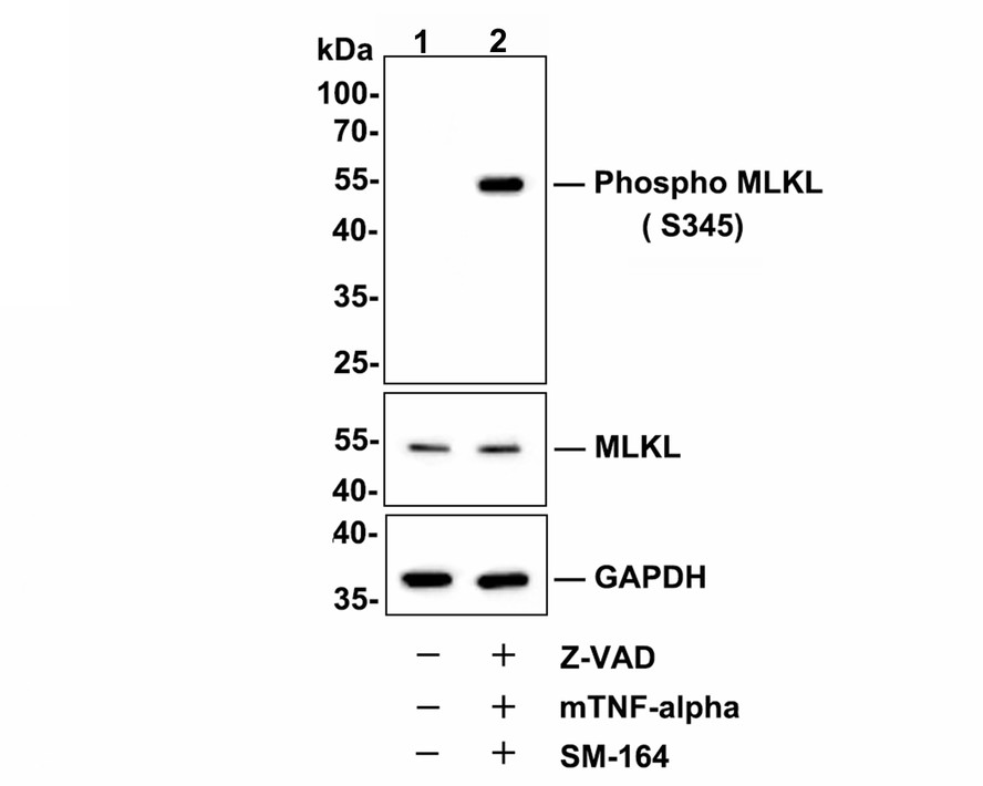 Western blot analysis of Phospho-MLKL (S345) on L929 cell lysates.Lane 1 : L929 cells, whole cell lysate, 10 μg /lane.Lane 2 : L929 cells were treated with 20 uM Z-VAD for 30 minutes, then added 20 ng/ml mTNF-alpha and 100 nM SM-164 for 4 hours, whole cell lysates, 10 μg/lane. 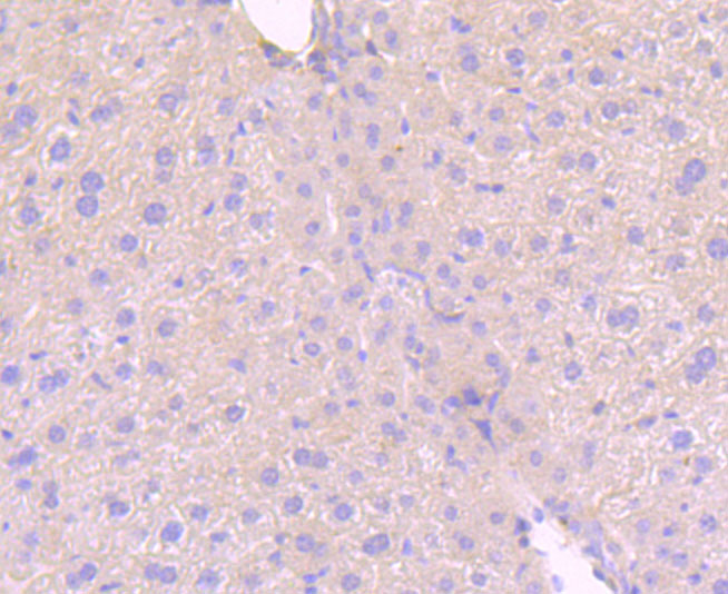 Immunohistochemical analysis of paraffin-embedded mouse liver tissue using anti-MLKL (phospho S345) antibody. The section was pre-treated using heat mediated antigen retrieval with Tris-EDTA buffer (pH 9.0) for 20 minutes.The tissues were blocked in 5% BSA for 30 minutes at room temperature, washed with ddH2O and PBS, and then probed with the primary antibody (bsm-54104R, 1/50) for 30 minutes at room temperature. The detection was performed using an HRP conjugated compact polymer system. DAB was used as the chromogen. Tissues were counterstained with hematoxylin and mounted with DPX.  Immunohistochemical analysis of paraffin-embedded mouse colon tissue using anti-MLKL (phospho S345) antibody. The section was pre-treated using heat mediated antigen retrieval with Tris-EDTA buffer (pH 9.0) for 20 minutes.The tissues were blocked in 5% BSA for 30 minutes at room temperature, washed with ddH2O and PBS, and then probed with the primary antibody (bsm-54104R, 1/50) for 30 minutes at room temperature. The detection was performed using an HRP conjugated compact polymer system. DAB was used as the chromogen. Tissues were counterstained with hematoxylin and mounted with DPX.  Immunohistochemical analysis of paraffin-embedded mouse spleen tissue using anti-MLKL (phospho S345) antibody. The section was pre-treated using heat mediated antigen retrieval with Tris-EDTA buffer (pH 9.0) for 20 minutes.The tissues were blocked in 5% BSA for 30 minutes at room temperature, washed with ddH2O and PBS, and then probed with the primary antibody (bsm-54104R, 1/50) for 30 minutes at room temperature. The detection was performed using an HRP conjugated compact polymer system. DAB was used as the chromogen. Tissues were counterstained with hematoxylin and mounted with DPX.  Immunohistochemical analysis of paraffin-embedded mouse lung tissue using anti-MLKL (phospho S345) antibody. The section was pre-treated using heat mediated antigen retrieval with Tris-EDTA buffer (pH 9.0) for 20 minutes.The tissues were blocked in 5% BSA for 30 minutes at room temperature, washed with ddH2O and PBS, and then probed with the primary antibody (bsm-54104R, 1/50) for 30 minutes at room temperature. The detection was performed using an HRP conjugated compact polymer system. DAB was used as the chromogen. Tissues were counterstained with hematoxylin and mounted with DPX.  Immunohistochemical analysis of paraffin-embedded mouse liver tissue using anti-Phospho-MLKL (S345) antibody. The section was pre-treated using heat mediated antigen retrieval with Tris-EDTA buffer (pH 9.0) for 20 minutes.The tissues were blocked in 5% BSA for 30 minutes at room temperature, washed with ddH2O and PBS, and then probed with the primary antibody (bsm-54104R, 1/50) for 30 minutes at room temperature. The detection was performed using an HRP conjugated compact polymer system. DAB was used as the chromogen. Tissues were counterstained with hematoxylin and mounted with DPX. 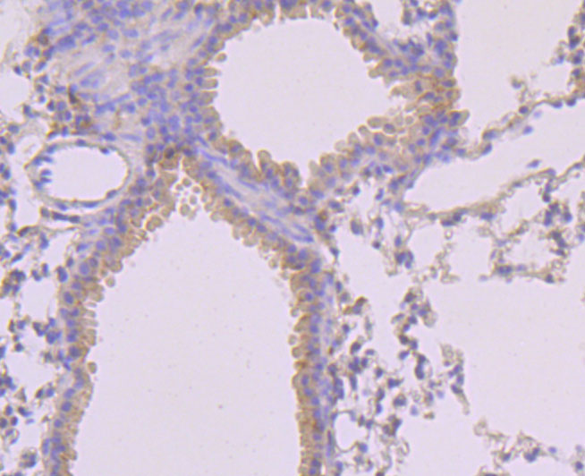 Immunohistochemical analysis of paraffin-embedded mouse lung tissue using anti-Phospho-MLKL (S345) antibody. The section was pre-treated using heat mediated antigen retrieval with Tris-EDTA buffer (pH 9.0) for 20 minutes.The tissues were blocked in 5% BSA for 30 minutes at room temperature, washed with ddH2O and PBS, and then probed with the primary antibody (bsm-54104R, 1/50) for 30 minutes at room temperature. The detection was performed using an HRP conjugated compact polymer system. DAB was used as the chromogen. Tissues were counterstained with hematoxylin and mounted with DPX. 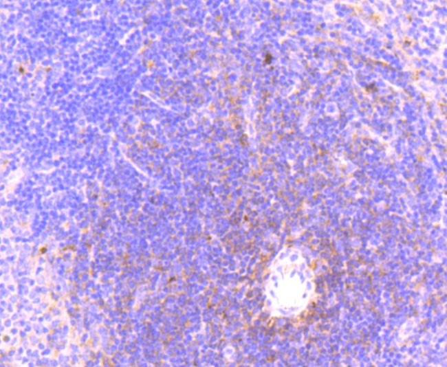 Immunohistochemical analysis of paraffin-embedded mouse spleen tissue using anti-Phospho-MLKL (S345) antibody. The section was pre-treated using heat mediated antigen retrieval with Tris-EDTA buffer (pH 9.0) for 20 minutes.The tissues were blocked in 5% BSA for 30 minutes at room temperature, washed with ddH2O and PBS, and then probed with the primary antibody (bsm-54104R, 1/50) for 30 minutes at room temperature. The detection was performed using an HRP conjugated compact polymer system. DAB was used as the chromogen. Tissues were counterstained with hematoxylin and mounted with DPX. 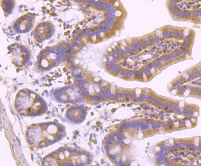 Immunohistochemical analysis of paraffin-embedded mouse colon tissue using anti-Phospho-MLKL (S345) antibody. The section was pre-treated using heat mediated antigen retrieval with Tris-EDTA buffer (pH 9.0) for 20 minutes.The tissues were blocked in 5% BSA for 30 minutes at room temperature, washed with ddH2O and PBS, and then probed with the primary antibody (bsm-54104R, 1/50) for 30 minutes at room temperature. The detection was performed using an HRP conjugated compact polymer system. DAB was used as the chromogen. Tissues were counterstained with hematoxylin and mounted with DPX. |


