
�Ϻ�ϸ����
��Դϸ��ϵ| ��תϸ��ϵ| �����ó���| �����ͻ��ϸ����| ���������ϸ����| ����ϸ��ϵ| ����ϸ��ϵ| ��ϸ��ϵ| �õ�ϸ��ϵ| Ȯ��ϸ��ϵ| ɽ���ϸ��ϵ| ���ϸ��ϵ| ���ϸ��ϵ| �����ϸ��ϵ| ����ϸ��ϵ| ţ��ϸ��| ����ϸ��ϵ| С��ϸ��ϵ| ����ϸ��ϵ|
��Դϸ��ϵ| ��תϸ��ϵ| �����ó���| �����ͻ��ϸ����| ���������ϸ����| ����ϸ��ϵ| ����ϸ��ϵ| ��ϸ��ϵ| �õ�ϸ��ϵ| Ȯ��ϸ��ϵ| ɽ���ϸ��ϵ| ���ϸ��ϵ| ���ϸ��ϵ| �����ϸ��ϵ| ����ϸ��ϵ| ţ��ϸ��| ����ϸ��ϵ| С��ϸ��ϵ| ����ϸ��ϵ|

| ��� | �۸� | ��� |
|---|---|---|
| 50ul | �� 1180.00 | 10 |
| 100ul | �� 1980.00 | 10 |
| ��Ʒ��� | bs-0081R |
| Ӣ������ | Rabbit?Anti-Caspase-3?antibody |
| �������� | ����װ��ᵰ��ø����-3���� |
| ��????�� | Caspase-3 subunit p17; cleaved Caspase 3; cleaved Caspase-3; APOPAIN; CASP3; Caspase 3 apoptosis related cysteine protease; Caspase3; CPP32; CPP32B; Cysteine protease CPP32; Human cysteine protease CPP32 isoform alpha mRNA complete cds; PARP cleavage protease; SCA 1; SCA1; SREBP cleavage activity 1; Yama; CASP3_HUMAN; Caspase-3; CASP-3; Apopain; Protein Yama; SREBP cleavage activity 1; SCA-1.?? |
 | Specific References??(269)?????|?????bs-0081R has been referenced in 269 publications. |
| �о����� | ����??ϸ������??������ѧ??�ź�ת��??ϸ������??Alzheimer's?? |
| ������Դ | Rabbit |
| ��¡���� | Polyclonal |
| ���淴Ӧ | Human,Mouse,Rat |
| ��ƷӦ�� | WB=1:500-2000, IHC-P=1:100-500, ICC=1:100, IF=1:100-500, ELISA=1:5000-10000 not yet tested in other applications. optimal dilutions/concentrations should be determined by the end user. |
| ���۷����� | 17/32kDa |
| ϸ����λ | ϸ����? |
| ��????״ | Liquid |
| Ũ????�� | 1mg/ml |
| ��?��?ԭ | KLH conjugated synthetic peptide derived from human caspase-3 p17 subunit:?80-175/277? |
| ��????�� | IgG |
| �������� | affinity purified by Protein A |
| ��?��?Һ | 0.01M TBS(pH7.4) with 1% BSA, 0.03% Proclin300 and 50% Glycerol. |
| �������� | Shipped at 4��. Store at -20 ��C for one year. Avoid repeated freeze/thaw cycles. |
| ע������ | This product as supplied is intended for research use only, not for use in human, therapeutic or diagnostic applications. |
| PubMed | PubMed |
| ��Ʒ���� | The caspase family of cysteine proteases play a key role in apoptosis. Caspase 3 is the most extensively studied apoptotic protein among caspase family members. Caspase 3 is synthesized as inactive pro enzyme that is processed in cells undergoing apoptosis by self proteolysis and/or cleavage by other upstream proteases (e.g. Caspases 8, 9 and 10). The processed form of Caspase 3 consists of large (17kDa) and small (12kDa) subunits which associate to form an active enzyme. Caspase 3 is cleaved at Asp28 Ser29 and Asp175 Ser176. The active Caspase 3 proteolytically cleaves and activates other caspases (e.g. Caspases 6, 7 and 9), as well as relevant targets in the cells (e.g. PARP and DFF). Alternative splicing of this gene results in two transcript variants which encode the same protein. In immunohistochemical studies Caspase 3 expression has been shown to be widespread but not present in all cell types (e.g. commonly reported in epithelial cells of skin, renal proximal tubules and collecting ducts). Differences in the level of Caspase 3 have been reported in cells of short lived nature (eg germinal centre B cells) and those that are long lived (eg mantle zone B cells). Caspase 3 is the predominant caspase involved in the cleavage of amyloid beta 4A precursor protein, which is associated with neuronal death in Alzheimer's disease. Reacts with Caspase-3 subunit p17 and precursor. Function: Involved in the activation cascade of caspases responsible for apoptosis execution. At the onset of apoptosis it proteolytically cleaves poly(ADP-ribose) polymerase (PARP) at a '216-Asp-|-Gly-217' bond. Cleaves and activates sterol regulatory element binding proteins (SREBPs) between the basic helix-loop-helix leucine zipper domain and the membrane attachment domain. Cleaves and activates caspase-6, -7 and -9. Involved in the cleavage of huntingtin. Triggers cell adhesion in sympathetic neurons through RET cleavage. Subunit: Heterotetramer that consists of two anti-parallel arranged heterodimers, each one formed by a 17 kDa (p17) and a 12 kDa (p12) subunit. Interacts with BIRC6/bruce. Subcellular Location: Cytoplasm. Tissue Specificity: Highly expressed in lung, spleen, heart, liver and kidney. Moderate levels in brain and skeletal muscle, and low in testis. Also found in many cell lines, highest expression in cells of the immune system. Post-translational modifications: Cleavage by granzyme B, caspase-6, caspase-8 and caspase-10 generates the two active subunits. Additional processing of the propeptides is likely due to the autocatalytic activity of the activated protease. Active heterodimers between the small subunit of caspase-7 protease and the large subunit of caspase-3 also occur and vice versa. S-nitrosylated on its catalytic site cysteine in unstimulated human cell lines and denitrosylated upon activation of the Fas apoptotic pathway, associated with an increase in intracellular caspase activity. Fas therefore activates caspase-3 not only by inducing the cleavage of the caspase zymogen to its active subunits, but also by stimulating the denitrosylation of its active site thiol. Similarity: Belongs to the peptidase C14A family. SWISS: P42574 Gene ID: 836 Database links: Entrez Gene: 836 Human Entrez Gene: 12367 Mouse Entrez Gene: 100008840 Rabbit Omim: 600636 Human SwissProt: P42574 Human SwissProt: P70677 Mouse SwissProt: Q8MJC3 Rabbit Unigene: 141125 Human Unigene: 34405 Mouse Unigene: 10562 Rat Caspase3�㷺�ֲ��ڸ��ֲ�ͬ���͵�ϸ���У���Caspase����������Ҫ�ĵ���ִ����֮һ�������Caspase-3��ʹ������ϸ���ṹ��ϸ�����ڼ�DNA������ص���øʧ��Ӷ�ʹϸ������. |
| ��ƷͼƬ | 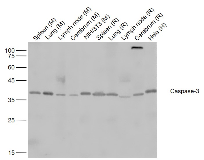 Sample: Lane 1: Spleen (Mouse) Lysate at 40 ug Lane 2: Lung (Mouse) Lysate at 40 ug Lane 3: Lymph node (Mouse) Lysate at 40 ug Lane 4: Cerebrum (Mouse) Lysate at 40 ug Lane 5: NIH/3T3 (Mouse) Cell Lysate at 30 ug Lane 6: Spleen (Rat) Lysate at 40 ug Lane 7: Lung (Rat) Lysate at 40 ug Lane 8: Lymph node (Rat) Lysate at 40 ug Lane 9: Cerebrum (Rat) Lysate at 40 ug Lane 10: Hela (Human) Cell Lysate at 30 ug Primary: Anti-Caspase-3 (bs-0081R) at 1/1000 dilution Secondary: IRDye800CW Goat Anti-Rabbit IgG at 1/20000 dilution Predicted band size: 35 kD Observed band size: 37 kD 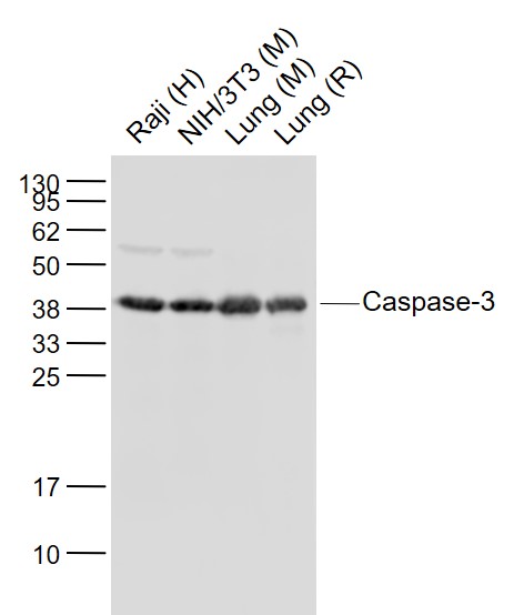 Sample: Lane 1: Raji (Human) Cell Lysate at 30 ug Lane 2: NIH/3T3 (Mouse) Cell Lysate at 30 ug Lane 3: Lung (Mouse) Lysate at 40 ug Lane 4: Lung (Rat) Lysate at 40 ug Primary: Anti-Caspase-3 (bs-0081R) at 1/1000 dilution Secondary: IRDye800CW Goat Anti-Rabbit IgG at 1/20000 dilution Predicted band size: 35 kD Observed band size: 37 kD 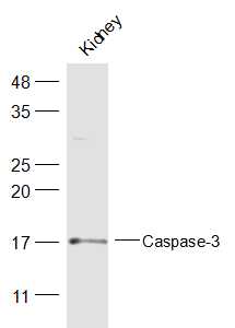 Sample: Kidney (Mouse) Lysate at 40 ug Primary: Anti-Caspase-3 (bs-0081R) at 1/300 dilution Secondary: IRDye800CW Goat Anti-Rabbit IgG at 1/20000 dilution Predicted band size: 28 kD Observed band size: 17 kD 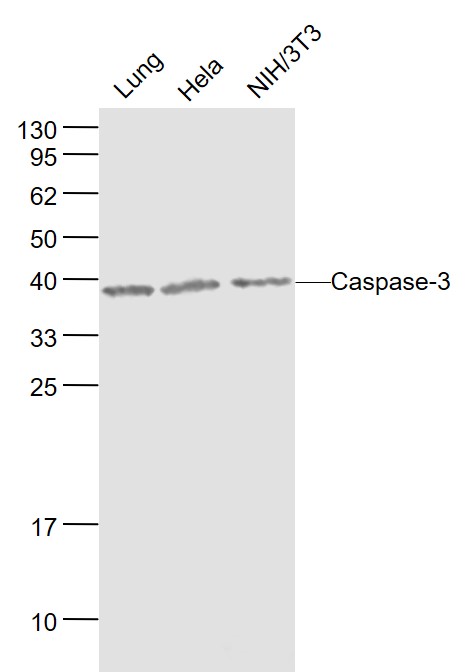 Sample: Lung(Mouse) Lysate at 40 ug Hela(Human) Cell Lysate at 30 ug NIH/3T3(Mouse) Cell Lysate at 30 ug Primary: Anti-Caspase-3 (bs-0081R) at 1/1000 dilution Secondary: IRDye800CW Goat Anti-Rabbit IgG at 1/20000 dilution Predicted band size: 35/29/19/17 kD Observed band size: 38 kD 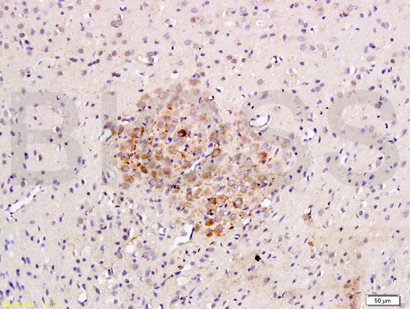 Tissue/cell: rat brain tissue; 4% Paraformaldehyde-fixed and paraffin-embedded; Antigen retrieval: citrate buffer ( 0.01M, pH 6.0 ), Boiling bathing for 15min; Block endogenous peroxidase by 3% Hydrogen peroxide for 30min; Blocking buffer (normal goat serum,C-0005) at 37�� for 20 min; Incubation: Anti-Caspase-3 Polyclonal Antibody, Unconjugated(bs-0081R) 1:200, overnight at 4�C, followed by conjugation to the secondary antibody(SP-0023) and DAB(C-0010) staining 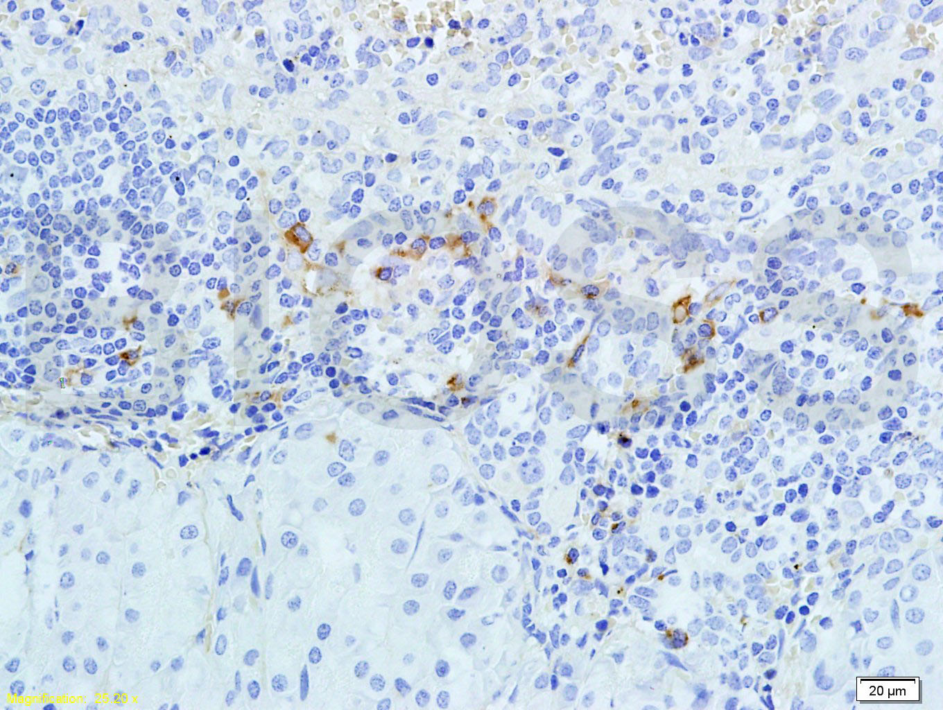 Tissue/cell: rabbit pancreas tissue; 4% Paraformaldehyde-fixed and paraffin-embedded; Antigen retrieval: citrate buffer ( 0.01M, pH 6.0 ), Boiling bathing for 15min; Block endogenous peroxidase by 3% Hydrogen peroxide for 30min; Blocking buffer (normal goat serum,C-0005) at 37�� for 20 min; Incubation: Anti-Caspase-3 Polyclonal Antibody, Unconjugated(bs-0081R) 1:300, overnight at 4�C, followed by conjugation to the secondary antibody(SP-0023) and DAB(C-0010) staining 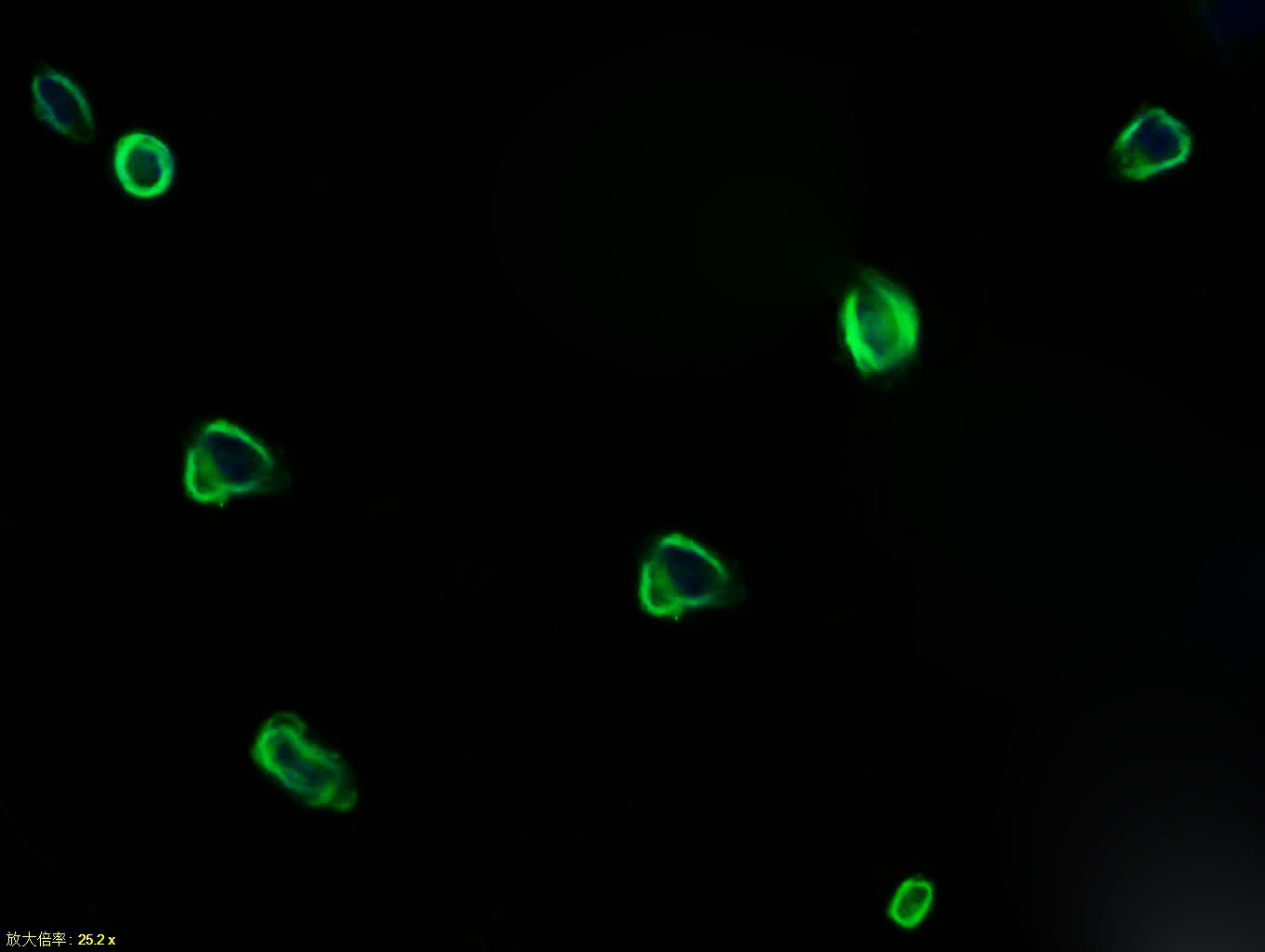 Tissue/cell: NIH/3T3 cell; 4% Paraformaldehyde-fixed; Triton X-100 at room temperature for 20 min; Blocking buffer (normal goat serum, C-0005) at 37��C for 20 min; Antibody incubation with (Caspase-3) polyclonal Antibody, Unconjugated (bs-0081R) 1:100, 90 minutes at 37��C; followed by a FITC conjugated Goat Anti-Rabbit IgG antibody at 37��C for 90 minutes, DAPI (blue, C02-04002) was used to stain the cell nuclei. |


