
上海细胞库
人源细胞系| 稳转细胞系| 基因敲除株| 基因点突变细胞株| 基因过表达细胞株| 重组细胞系| 猪的细胞系| 马细胞系| 兔的细胞系| 犬的细胞系| 山羊的细胞系| 鱼的细胞系| 猴的细胞系| 仓鼠的细胞系| 狗的细胞系| 牛的细胞| 大鼠细胞系| 小鼠细胞系| 其他细胞系|
人源细胞系| 稳转细胞系| 基因敲除株| 基因点突变细胞株| 基因过表达细胞株| 重组细胞系| 猪的细胞系| 马细胞系| 兔的细胞系| 犬的细胞系| 山羊的细胞系| 鱼的细胞系| 猴的细胞系| 仓鼠的细胞系| 狗的细胞系| 牛的细胞| 大鼠细胞系| 小鼠细胞系| 其他细胞系|

| 规格 | 价格 | 库存 |
|---|---|---|
| 50ul | ¥ 1180.00 | 10 |
| 100ul | ¥ 1980.00 | 10 |
| 200ul | ¥ 2800.00 | 10 |
| 产品编号 | bs-5220R |
| 英文名称 | Rabbit?Anti-Phospho-Bcl-2 (Thr129)?antibody |
| 中文名称 | 磷酸化Bcl-2抗体 |
| 别????名 | Bcl-2 (phospho T129); Bcl-2 (phospho Thr129); p-Bcl-2 (Thr129); Apoptosis regulator Bcl 2; Apoptosis regulator Bcl2; AW986256; B cell CLL/lymphoma 2; B cell leukemia/lymphoma 2; B cell lymphoma 2; Bcl 2; Bcl-2; Bcl2; BCL2 protein; C430015F12Rik; D630044D05Rik; D830018M01Rik; Leukemia/lymphoma, B-cell, 2; Oncogene B-cell leukemia 2; BCL2_HUMAN.?? |
| 产品类型 | 磷酸化抗体? |
| 研究领域 | 肿瘤??细胞生物??免疫学??神经生物学??信号转导??细胞凋亡??转录调节因子??线粒体?? |
| 抗体来源 | Rabbit |
| 克隆类型 | Polyclonal |
| 交叉反应 | Human,Mouse,Rat (predicted: Dog,Pig,Cow,Horse,Sheep) |
| 产品应用 | WB=1:500-2000, IHC-P=1:100-500, IHC-F=1:100-500, IF=1:100-500, Flow-Cyt=1μg/Test, ELISA=1:5000-10000 not yet tested in other applications. optimal dilutions/concentrations should be determined by the end user. |
| 理论分子量 | 26kDa |
| 细胞定位 | 细胞核?细胞浆?细胞膜?线粒体 |
| 性????状 | Liquid |
| 浓????度 | 1mg/ml |
| 免?疫?原 | KLH conjugated Synthesised phosphopeptide derived from human Bcl-2 around the phosphorylation site of Thr129:?FA(p-T)VV? |
| 亚????型 | IgG |
| 纯化方法 | affinity purified by Protein A |
| 缓?冲?液 | 0.01M TBS(pH7.4) with 1% BSA, 0.03% Proclin300 and 50% Glycerol. |
| 保存条件 | Shipped at 4℃. Store at -20 °C for one year. Avoid repeated freeze/thaw cycles. |
| 注意事项 | This product as supplied is intended for research use only, not for use in human, therapeutic or diagnostic applications. |
| PubMed | PubMed |
| 产品介绍 | BCL2 is an integral outer mitochondrial membrane protein that blocks the apoptotic death of some cells such as lymphocytes. Constitutive expression of BCL2, such as in the case of translocation of BCL2 to Ig heavy chain locus, is thought to be the cause of follicular lymphoma. Two transcript variants (alpha and beta) produced by alternate splicing, differ in their C-terminal ends. BCL2 suppresses apoptosis in a variety of cell systems including factor-dependent lymphohematopoietic and neural cells. It regulates cell death by controlling the mitochondrial membrane permeability. It appears to function in a feedback loop system with caspases. BCL2 inhibits caspase activity either by preventing the release of cytochrome c from the mitochondria and/or by binding to the apoptosis-activating factor (APAF1). It can form homodimers, and heterodimers with BAX, BAD, BAK and BclX(L). Heterodimerization with BAX requires intact BH1 and BH2 domains, and is necessary for anti-apoptotic activity. Function: Suppresses apoptosis in a variety of cell systems including factor-dependent lymphohematopoietic and neural cells. Regulates cell death by controlling the mitochondrial membrane permeability. Appears to function in a feedback loop system with caspases. Inhibits caspase activity either by preventing the release of cytochrome c from the mitochondria and/or by binding to the apoptosis-activating factor (APAF-1). Subunit: Forms homodimers, and heterodimers with BAX, BAD, BAK and Bcl-X(L). Heterodimerization with BAX requires intact BH1 and BH2 motifs, and is necessary for anti-apoptotic activity. Interacts with EI24 (By similarity). Also interacts with APAF1, BBC3, BCL2L1, BNIPL, MRPL41 and TP53BP2. Binding to FKBP8 seems to target BCL2 to the mitochondria and probably interferes with the binding of BCL2 to its targets. Interacts with BAG1 in an ATP-dependent manner. Interacts with RAF1 (the 'Ser-338' and 'Ser-339' phosphorylated form). Interacts (via the BH4 domain) with EGLN3; the interaction prevents the formation of the BAX-BCL2 complex and inhibits the anti-apoptotic activity of BCL2. Interacts with G0S2; this interaction also prevents the formation of the anti-apoptotic BAX-BCL2 complex. Subcellular Location: Mitochondrion outer membrane; Single-pass membrane protein. Nucleus membrane; Single-pass membrane protein. Endoplasmic reticulum membrane; Single-pass membrane protein. Tissue Specificity: Expressed in a variety of tissues. Post-translational modifications: Phosphorylation/dephosphorylation on Ser-70 regulates anti-apoptotic activity. Growth factor-stimulated phosphorylation on Ser-70 by PKC is required for the anti-apoptosis activity and occurs during the G2/M phase of the cell cycle. In the absence of growth factors, BCL2 appears to be phosphorylated by other protein kinases such as ERKs and stress-activated kinases. Phosphorylated by MAPK8/JNK1 at Thr-69, Ser-70 and Ser-87, wich stimulates starvation-induced autophagy. Dephosphorylated by protein phosphatase 2A (PP2A). Proteolytically cleaved by caspases during apoptosis. The cleaved protein, lacking the BH4 motif, has pro-apoptotic activity, causes the release of cytochrome c into the cytosol promoting further caspase activity. Monoubiquitinated by PARK2, leading to increase its stability. DISEASE: Note=A chromosomal aberration involving BCL2 has been found in chronic lymphatic leukemia. Translocation t(14;18)(q32;q21) with immunoglobulin gene regions. BCL2 mutations found in non-Hodgkin lymphomas carrying the chromosomal translocation could be attributed to the Ig somatic hypermutation mechanism resulting in nucleotide transitions. Similarity: Belongs to the Bcl-2 family. SWISS: P49950 Gene ID: 596 Database links: Entrez Gene: 596 Human Entrez Gene: 12043 Mouse Omim: 151430 Human SwissProt: P10415 Human SwissProt: P10417 Mouse Unigene: 150749 Human Unigene: 257460 Mouse Unigene: 9996 Rat |
| 产品图片 | 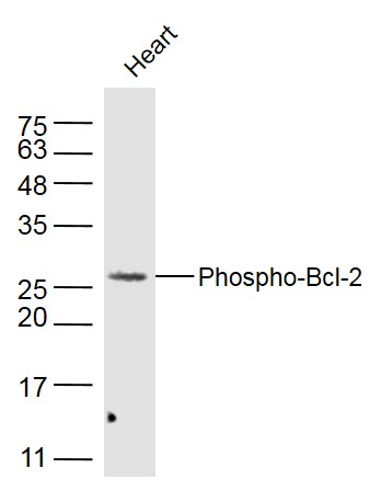 Sample: Heart(Mouse) Lysate at 40 ug Primary: Anti-Phospho-Bcl-2 (Thr129) (bs-5220R) at 1/300 dilution Secondary: IRDye800CW Goat Anti-Rabbit IgG at 1/20000 dilution Predicted band size: 26 kD Observed band size: 26 kD 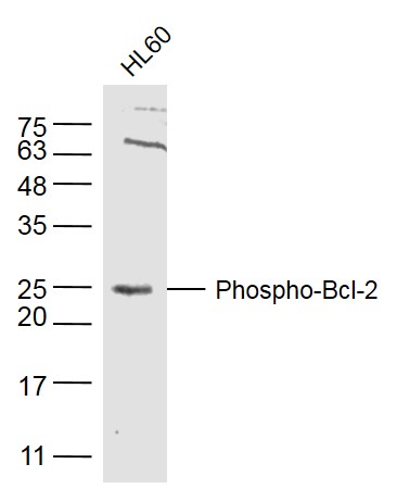 Sample: HL60(Human) Cell Lysate at 40 ug Primary: Anti-Phospho-Bcl-2 (Thr129) (bs-5220R) at 1/300 dilution Secondary: IRDye800CW Goat Anti-Rabbit IgG at 1/20000 dilution Predicted band size: 26 kD Observed band size: 26 kD 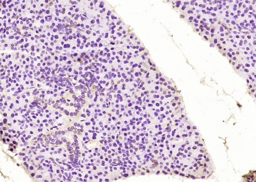 Paraformaldehyde-fixed, paraffin embedded (mouse lymphoid); Antigen retrieval by boiling in sodium citrate buffer (pH6.0) for 15min; Block endogenous peroxidase by 3% hydrogen peroxide for 20 minutes; Blocking buffer (normal goat serum) at 37°C for 30min; Antibody incubation with (Phospho-Bcl-2 (Thr129)) Polyclonal Antibody, Unconjugated (bs-5220R) at 1:200 overnight at 4°C, followed by operating according to SP Kit(Rabbit) (sp-0023) instructionsand DAB staining. 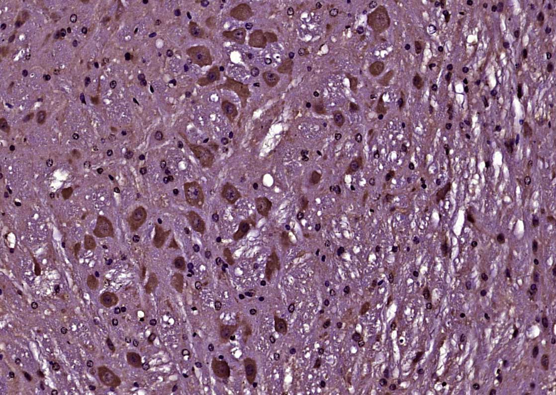 Paraformaldehyde-fixed, paraffin embedded (Mouse brain); Antigen retrieval by boiling in sodium citrate buffer (pH6.0) for 15min; Block endogenous peroxidase by 3% hydrogen peroxide for 20 minutes; Blocking buffer (normal goat serum) at 37°C for 30min; Antibody incubation with (Phospho-Bcl-2 (Thr129)) Polyclonal Antibody, Unconjugated (bs-5220R) at 1:400 overnight at 4°C, followed by operating according to SP Kit(Rabbit) (sp-0023) instructionsand DAB staining. 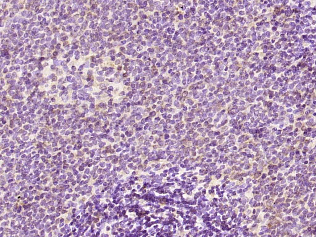 Paraformaldehyde-fixed, paraffin embedded (rat spleen); Antigen retrieval by boiling in sodium citrate buffer (pH6.0) for 15min; Block endogenous peroxidase by 3% hydrogen peroxide for 20 minutes; Blocking buffer (normal goat serum) at 37°C for 30min; Antibody incubation with (Phospho-Bcl-2 (Thr129)) Polyclonal Antibody, Unconjugated (bs-5220R) at 1:200 overnight at 4°C, followed by operating according to SP Kit(Rabbit) (sp-0023) instructionsand DAB staining. 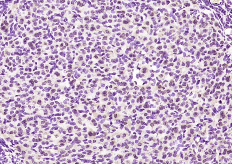 Paraformaldehyde-fixed, paraffin embedded (mouse ovary); Antigen retrieval by boiling in sodium citrate buffer (pH6.0) for 15min; Block endogenous peroxidase by 3% hydrogen peroxide for 20 minutes; Blocking buffer (normal goat serum) at 37°C for 30min; Antibody incubation with (Phospho-Bcl-2 (Thr129)) Polyclonal Antibody, Unconjugated (bs-5220R) at 1:200 overnight at 4°C, followed by operating according to SP Kit(Rabbit) (sp-0023) instructionsand DAB staining. 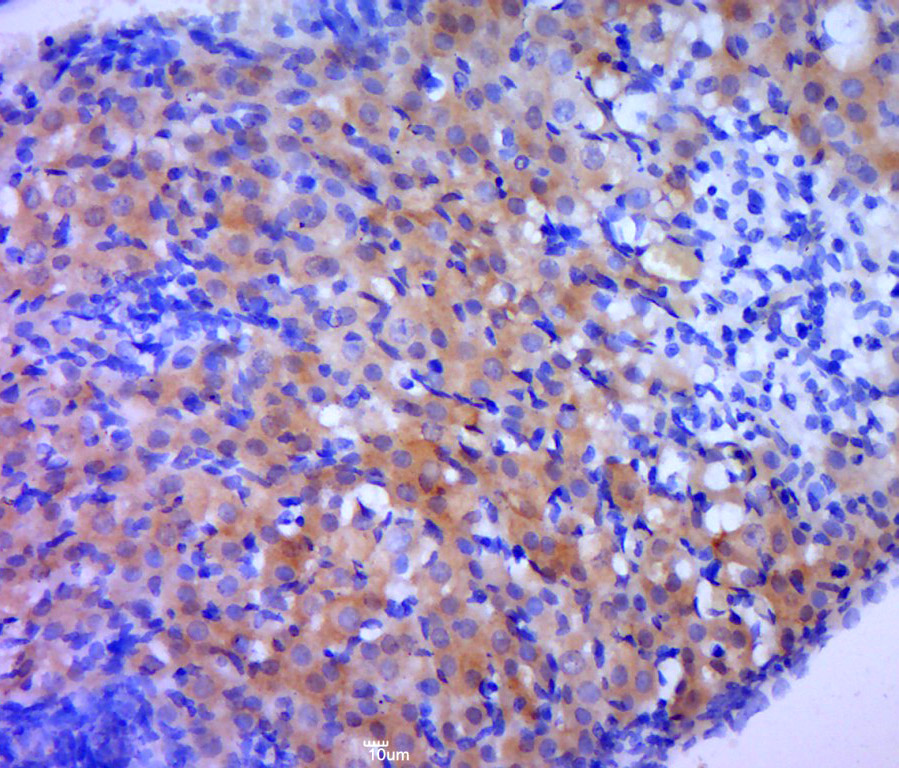 Paraformaldehyde-fixed, paraffin embedded (rat ovary tissue); Antigen retrieval by boiling in sodium citrate buffer (pH6.0) for 15min; Block endogenous peroxidase by 3% hydrogen peroxide for 20 minutes; Blocking buffer (normal goat serum) at 37°C for 30min; Antibody incubation with (P-Bcl-2(Thr129)) Polyclonal Antibody, Unconjugated (bs-5220R) at 1:400 overnight at 4°C, followed by a conjugated secondary (sp-0023) for 20 minutes and DAB staining. 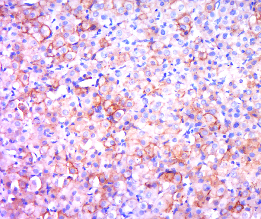 Paraformaldehyde-fixed, paraffin embedded (rat ovary); Antigen retrieval by boiling in sodium citrate buffer (pH6.0) for 15min; Block endogenous peroxidase by 3% hydrogen peroxide for 20 minutes; Blocking buffer (normal goat serum) at 37°C for 30min; Antibody incubation with (BCL2) Polyclonal Antibody, Unconjugated (bs-5220R) at 1:400 overnight at 4°C, followed by a conjugated secondary (sp-0023) for 20 minutes and DAB staining. 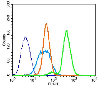 Blank control(blue):K562 (fixed with 2% paraformaldehyde for 10 min at 37℃). Primary Antibody:Rabbit Anti-Phospho-Bcl-2(Thr129)antibody (bs-5220R,Green); Dilution: 1μg in 100 μL 1X PBS containing 0.5% BSA; Isotype Control Antibody: Rabbit IgG(orange),used under the same conditions; Secondary Antibody: Goat anti-rabbit IgG-FITC(white blue), Dilution: 1:200 in 1 X PBS containing 0.5% BSA. 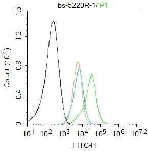 Blank control:HL-60. Primary Antibody (green line): Rabbit Anti-Phospho-Bcl-2 (Thr129) antibody (bs-5220R) Dilution: 1μg /10^6 cells; Isotype Control Antibody (orange line): Rabbit IgG . Secondary Antibody : Goat anti-rabbit IgG-AF488 Dilution: 1μg /test. Protocol The cells were fixed with 4% PFA (10min at room temperature)and then permeabilized with 0.1%PBST for 20 min at room temperature. The cells were then incubated in 5%BSA to block non-specific protein-protein interactions for 30 min at room temperature .Cells stained with Primary Antibody for 30 min at room temperature. The secondary antibody used for 40 min at room temperature. Acquisition of 20,000 events was performed. 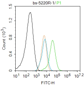 Blank control:HL-60. Primary Antibody (green line): Rabbit Anti-Phospho-Bcl-2 (Thr129) antibody (bs-5220R) Dilution: 1μg /10^6 cells; Isotype Control Antibody (orange line): Rabbit IgG . Secondary Antibody : Goat anti-rabbit IgG-AF488 Dilution: 1μg /test. Protocol The cells were fixed with 4% PFA (10min at room temperature)and then permeabilized with 0.1%PBST for 20 min at room temperature. The cells were then incubated in 5%BSA to block non-specific protein-protein interactions for 30 min at room temperature .Cells stained with Primary Antibody for 30 min at room temperature. The secondary antibody used for 40 min at room temperature. Acquisition of 20,000 events was performed. |


