
上海细胞库
人源细胞系| 稳转细胞系| 基因敲除株| 基因点突变细胞株| 基因过表达细胞株| 重组细胞系| 猪的细胞系| 马细胞系| 兔的细胞系| 犬的细胞系| 山羊的细胞系| 鱼的细胞系| 猴的细胞系| 仓鼠的细胞系| 狗的细胞系| 牛的细胞| 大鼠细胞系| 小鼠细胞系| 其他细胞系|
人源细胞系| 稳转细胞系| 基因敲除株| 基因点突变细胞株| 基因过表达细胞株| 重组细胞系| 猪的细胞系| 马细胞系| 兔的细胞系| 犬的细胞系| 山羊的细胞系| 鱼的细胞系| 猴的细胞系| 仓鼠的细胞系| 狗的细胞系| 牛的细胞| 大鼠细胞系| 小鼠细胞系| 其他细胞系|

| 规格 | 价格 | 库存 |
|---|---|---|
| 50ul | ¥ 1580.00 | 10 |
| 100ul | ¥ 2500.00 | 10 |
| 产品编号 | bsm-60235R |
| 英文名称 | Rabbit?Anti-Keratin 6?antibody |
| 中文名称 | 细胞角蛋白6兔单克隆抗体 |
| 别????名 | Keratin, type II cytoskeletal 6C; KRT6C; Cytokeratin-6C; CK-6C; Cytokeratin-6E; CK-6E; Keratin K6h; Keratin-6C; K6C; Type-II keratin Kb12; KRT6E; K2C6C_HUMAN?? |
| 研究领域 | 肿瘤??信号转导?? |
| 抗体来源 | Rabbit |
| 克隆类型 | Recombinant |
| 克?隆?号 | B12C5 |
| 交叉反应 | Human,Mouse,Rat |
| 产品应用 | WB=1:500-2000, IHC-P=1:100-500, ICC=1:20-100, IF=1:50 not yet tested in other applications. optimal dilutions/concentrations should be determined by the end user. |
| 理论分子量 | 60kDa |
| 细胞定位 | 细胞浆? |
| 性????状 | Liquid |
| 浓????度 | 1mg/ml |
| 免?疫?原 | KLH conjugated synthetic peptide derived from human Keratin 6? |
| 亚????型 | IgG |
| 纯化方法 | affinity purified by Protein A |
| 缓?冲?液 | 0.01M TBS(pH7.4) with 1% BSA, 0.03% Proclin300 and 50% Glycerol. |
| 保存条件 | Shipped at 4℃. Store at -20 °C for one year. Avoid repeated freeze/thaw cycles. |
| 注意事项 | This product as supplied is intended for research use only, not for use in human, therapeutic or diagnostic applications. |
| PubMed | PubMed |
| 产品介绍 | Keratins are intermediate filament proteins responsible for the structural integrity of epithelial cells and are subdivided into epithelial keratins and hair keratins. The type II keratins are clustered in a region of chromosome 12q13. [provided by RefSeq, Jul 2009] Function: There are at least six isoforms of human type II keratin-6 (K6). There are two types of cytoskeletal and microfibrillar keratin, I (acidic) and II (neutral to basic) (40-55 and 56-70 kDa, respectively). Subunit: Heterodimer of a type I and a type II keratin. KRT6 isomers associate with KRT16 and/or KRT17. Interacts with TCHP. Tissue Specificity: Constitutively expressed in distinct types of epithelia such as those in oral mucosa, esophagus, papillae of tongue and hair follicle outer root sheath. DISEASE: Defects in KRT6A are a cause of pachyonychia congenital type 1 (PC1) [MIM:167200]; also known as Jadassohn-Lewandowsky syndrome. PC1 is an autosomal dominant ectodermal dysplasia characterized by hypertrophic nail dystrophy resulting in onchyogryposis (thickening and increase in curvature of the nail), palmoplantar keratoderma, follicular hyperkeratosis, and oral leukokeratosis. Hyperhidrosis of the hands and feet is usually present. Similarity: Belongs to the intermediate filament family. SWISS: P48668 Gene ID: 286887 Database links: Entrez Gene: 286887?Human Entrez Gene: 16687?Mouse SwissProt: P48668?Human SwissProt: P50446?Mouse |
| 产品图片 | 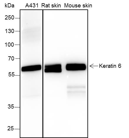 Blocking buffer: 5% NFDM/TBST Primary ab dilution: 1:2000 Primary ab incubation condition: 2 hours at room temperature Lysate: A431, Rat skin, Mouse skin Protein loading quantity: 20 μg Exposure time: 15 s Predicted MW: 60 kDa Observed MW: 56 kDa 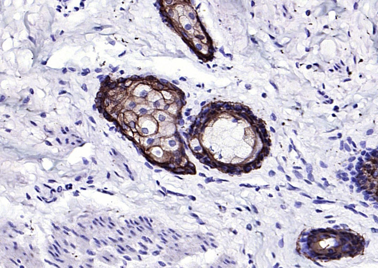 Paraformaldehyde-fixed, paraffin embedded (human breast); Antigen retrieval by boiling in sodium citrate buffer (pH6.0) for 15min; Block endogenous peroxidase by 3% hydrogen peroxide for 20 minutes; Blocking buffer (normal goat serum) at 37°C for 30min; Antibody incubation with (Keratin 6) Monoclonal Antibody, Unconjugated (bsm-60235R) at 1:200 overnight at 4°C, followed by operating according to SP Kit(Rabbit) (sp-0023) instructionsand DAB staining. 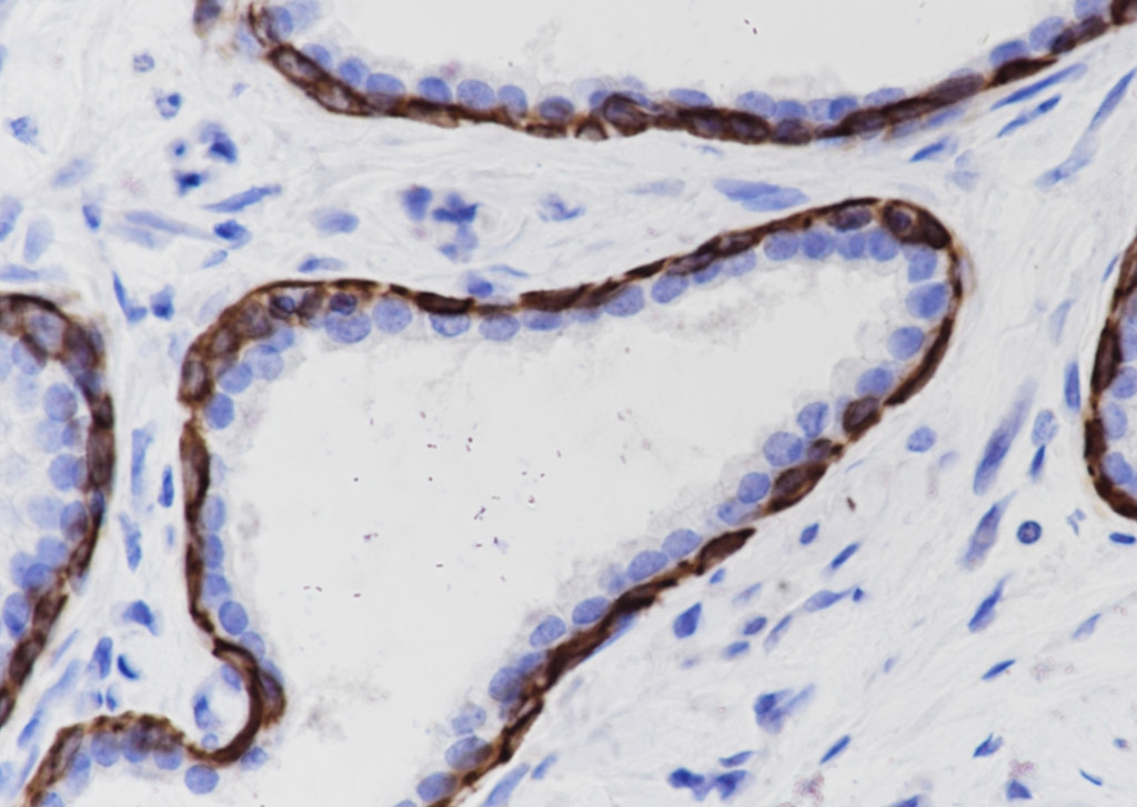 Tissue: Human prostate hyperplasia Section type: Formalin fixed & Paraffin -embedded section Retrieval method: High temperature and high pressure Retrieval buffer: Tris/EDTA buffer, pH 9.0 Primary ab dilution: 1:1000 Primary ab incubation condition: 1 hour at room temperature Counter stain: Hematoxylin Comment: Color brown is the positive signal for bsm-60235R 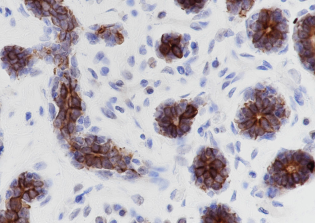 Tissue: Human breast Section type: Formalin fixed & Paraffin -embedded section Retrieval method: High temperature and high pressure Retrieval buffer: Tris/EDTA buffer, pH 9.0 Primary ab dilution: 1:1000 Primary ab incubation condition: 1 hour at room temperature Counter stain: Hematoxylin Comment: Color brown is the positive signal for bsm-60235R 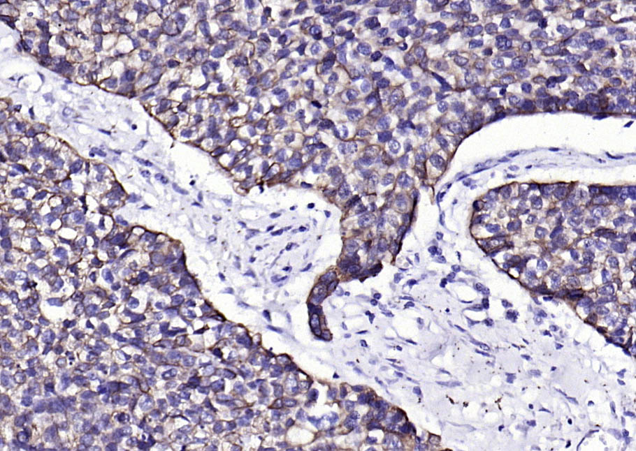 Paraformaldehyde-fixed, paraffin embedded (human esophageal); Antigen retrieval by boiling in sodium citrate buffer (pH6.0) for 15min; Block endogenous peroxidase by 3% hydrogen peroxide for 20 minutes; Blocking buffer (normal goat serum) at 37°C for 30min; Antibody incubation with (Keratin 6) Monoclonal Antibody, Unconjugated (bsm-60235R) at 1:200 overnight at 4°C, followed by operating according to SP Kit(Rabbit) (sp-0023) instructionsand DAB staining.  Paraformaldehyde-fixed, paraffin embedded (mouse esophageal); Antigen retrieval by boiling in sodium citrate buffer (pH6.0) for 15min; Block endogenous peroxidase by 3% hydrogen peroxide for 20 minutes; Blocking buffer (normal goat serum) at 37°C for 30min; Antibody incubation with (Keratin 6) Monoclonal Antibody, Unconjugated (bsm-60235R) at 1:200 overnight at 4°C, followed by operating according to SP Kit(Rabbit) (sp-0023) instructionsand DAB staining. 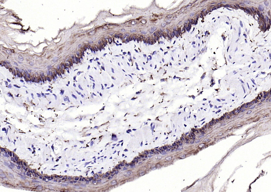 Paraformaldehyde-fixed, paraffin embedded (rat esophageal); Antigen retrieval by boiling in sodium citrate buffer (pH6.0) for 15min; Block endogenous peroxidase by 3% hydrogen peroxide for 20 minutes; Blocking buffer (normal goat serum) at 37°C for 30min; Antibody incubation with (Keratin 6) Monoclonal Antibody, Unconjugated (bsm-60235R) at 1:200 overnight at 4°C, followed by operating according to SP Kit(Rabbit) (sp-0023) instructionsand DAB staining. 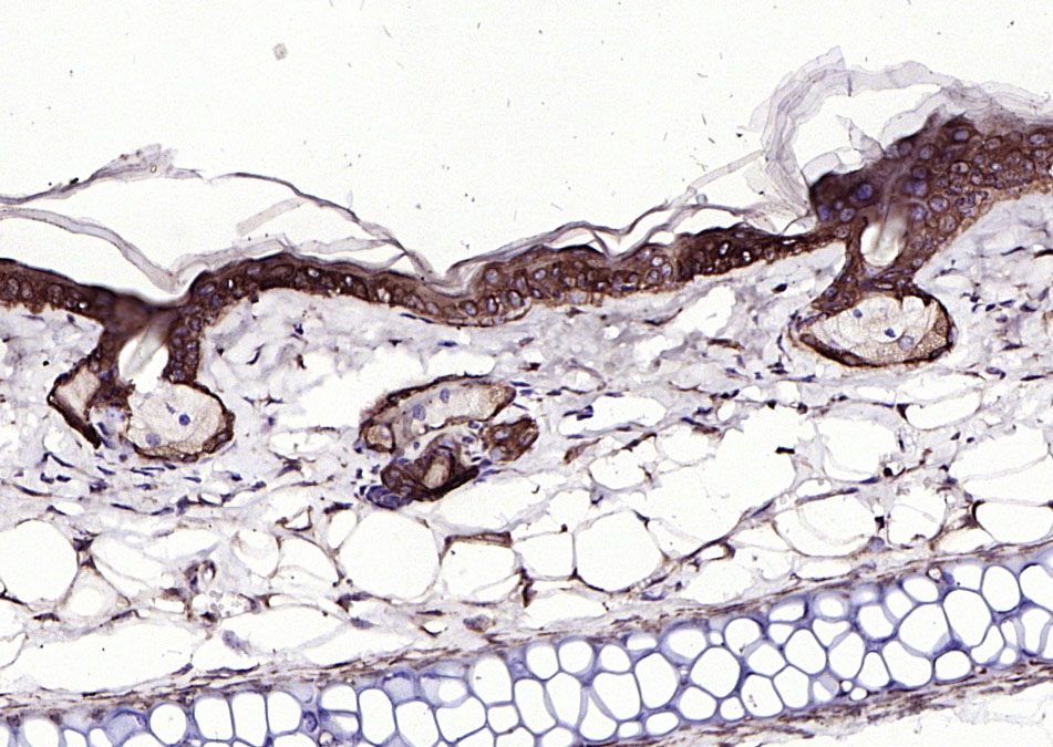 Paraformaldehyde-fixed, paraffin embedded (mouse skin); Antigen retrieval by boiling in sodium citrate buffer (pH6.0) for 15min; Block endogenous peroxidase by 3% hydrogen peroxide for 20 minutes; Blocking buffer (normal goat serum) at 37°C for 30min; Antibody incubation with (Keratin 6) Monoclonal Antibody, Unconjugated (bsm-60235R) at 1:200 overnight at 4°C, followed by operating according to SP Kit(Rabbit) (sp-0023) instructionsand DAB staining. 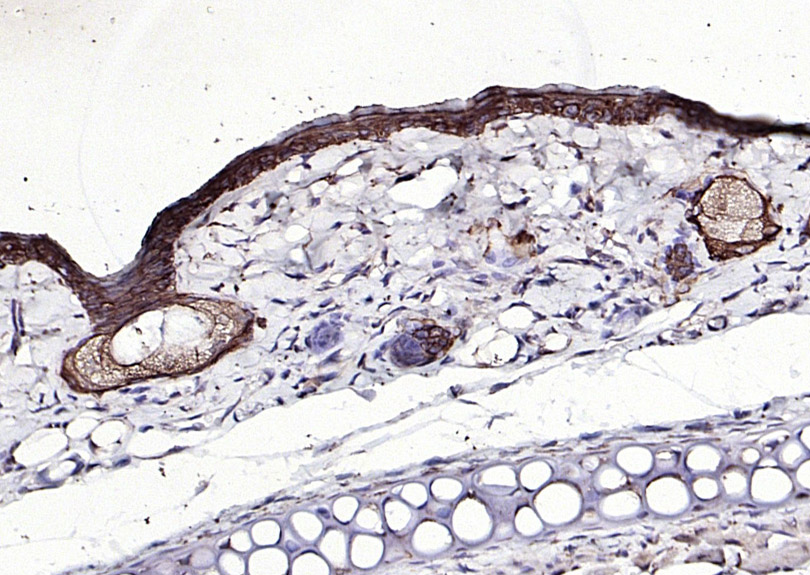 Paraformaldehyde-fixed, paraffin embedded (rat skin); Antigen retrieval by boiling in sodium citrate buffer (pH6.0) for 15min; Block endogenous peroxidase by 3% hydrogen peroxide for 20 minutes; Blocking buffer (normal goat serum) at 37°C for 30min; Antibody incubation with (Keratin 6) Monoclonal Antibody, Unconjugated (bsm-60235R) at 1:200 overnight at 4°C, followed by operating according to SP Kit(Rabbit) (sp-0023) instructionsand DAB staining. 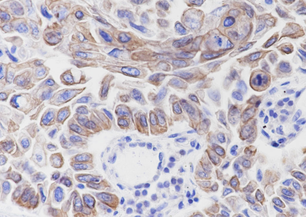 Tissue: Human lung squamous cell carcinoma Section type: Formalin fixed & Paraffin -embedded section Retrieval method: High temperature and high pressure Retrieval buffer: Tris/EDTA buffer, pH 9.0 Primary ab dilution: 1:1000 Primary incubation condition: 1 hour at room temperature Counter stain: Hematoxylin Comment: Color brown is the positive signal for bsm-60235R 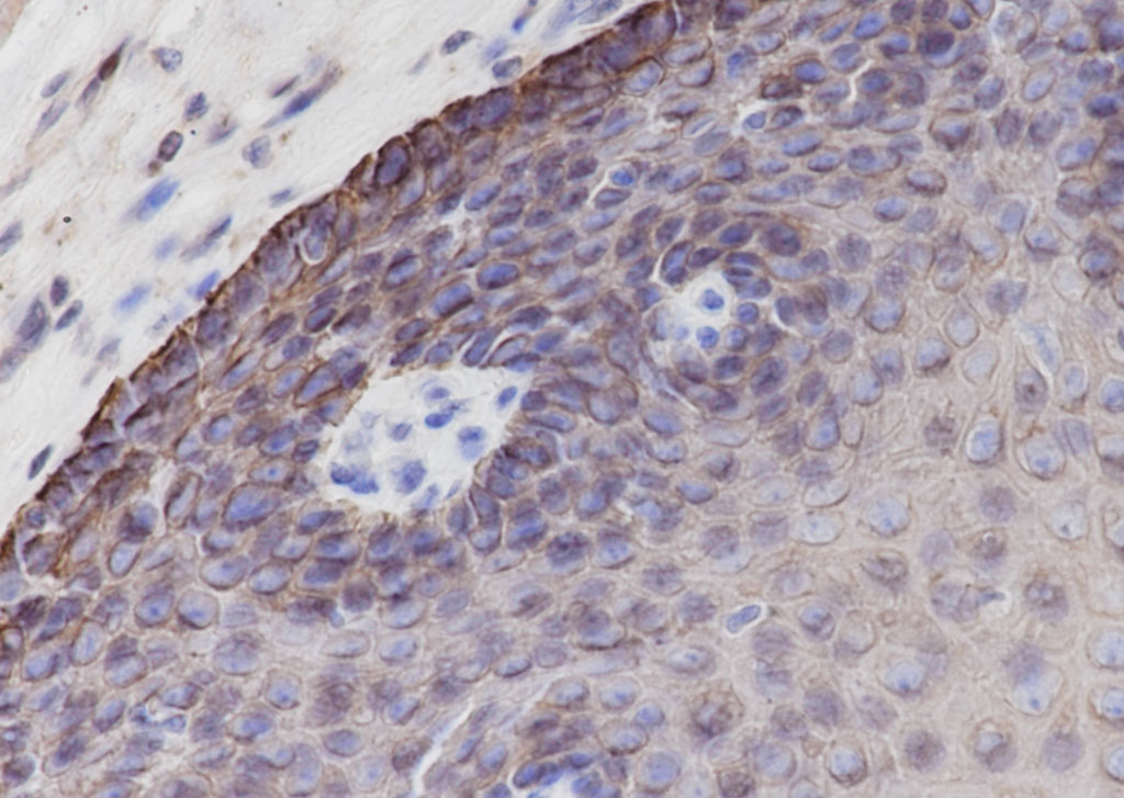 Tissue: Human esophagus Section type: Formalin fixed & Paraffin -embedded section Retrieval method: High temperature and high pressure Retrieval buffer: Tris/EDTA buffer, pH 9.0 Primary ab dilution: 1:1000 Primary incubation condition: 1 hour at room temperature Counter stain: Hematoxylin Comment: Color brown is the positive signal for bsm-60235R  Cell line: HaCat Fixative: 4% Paraformaldehyde Permeabilization: 0.1% TritonX-100 Primary ab dilution: 1:50 Primary incubation condition: 4°C overnight Nuclear counter stain: DAPI (Blue) Comment: Color green is the positive signal for bsm-60235R |


