
�Ϻ�ϸ����
��Դϸ��ϵ| ��תϸ��ϵ| �����ó���| �����ͻ��ϸ����| ���������ϸ����| ����ϸ��ϵ| ����ϸ��ϵ| ��ϸ��ϵ| �õ�ϸ��ϵ| Ȯ��ϸ��ϵ| ɽ���ϸ��ϵ| ���ϸ��ϵ| ���ϸ��ϵ| �����ϸ��ϵ| ����ϸ��ϵ| ţ��ϸ��| ����ϸ��ϵ| С��ϸ��ϵ| ����ϸ��ϵ|
��Դϸ��ϵ| ��תϸ��ϵ| �����ó���| �����ͻ��ϸ����| ���������ϸ����| ����ϸ��ϵ| ����ϸ��ϵ| ��ϸ��ϵ| �õ�ϸ��ϵ| Ȯ��ϸ��ϵ| ɽ���ϸ��ϵ| ���ϸ��ϵ| ���ϸ��ϵ| �����ϸ��ϵ| ����ϸ��ϵ| ţ��ϸ��| ����ϸ��ϵ| С��ϸ��ϵ| ����ϸ��ϵ|

| ��� | �۸� | ��� |
|---|---|---|
| 50ul | �� 1180.00 | 10 |
| 100ul | �� 1980.00 | 10 |
| 200ul | �� 2800.00 | 10 |
| ��Ʒ��� | bs-0199R |
| Ӣ������ | Rabbit?Anti-GFAP?antibody |
| �������� | ������ά���Ե����� |
| ��????�� | Astrocyte Marker; FLJ45472; GFAP; Glial Fibrillary Acidic Protein; Intermediate filament protein; GFAP_HUMAN.?? |
 | Specific References??(78)?????|?????bs-0199R has been referenced in 78 publications. |
| �о����� | ����??ϸ������??����ѧ??������ѧ??�ź�ת��??��ϸ��??ϸ��ճ������??ϸ�����ͱ�־��??ϸ���Ǽ�?? |
| ������Դ | Rabbit |
| ��¡���� | Polyclonal |
| ���淴Ӧ | Human,Mouse,Rat (predicted: Dog,Pig,Cow,Rabbit,Sheep) |
| ��ƷӦ�� | WB=1:500-2000, IHC-P=1:200-1000, IHC-F=1:200-1000, IF=1:200-800, Flow-Cyt=1��g/Test, ELISA=1:5000-10000 not yet tested in other applications. optimal dilutions/concentrations should be determined by the end user. |
| ���۷����� | 48kDa |
| ϸ����λ | ϸ����? |
| ��????״ | Liquid |
| Ũ????�� | 1mg/ml |
| ��?��?ԭ | KLH conjugated synthetic peptide derived from human GFAP:?51-150/432? |
| ��????�� | IgG |
| �������� | affinity purified by Protein A |
| ��?��?Һ | 0.01M TBS(pH7.4) with 1% BSA, 0.03% Proclin300 and 50% Glycerol. |
| �������� | Shipped at 4��. Store at -20 ��C for one year. Avoid repeated freeze/thaw cycles. |
| ע������ | This product as supplied is intended for research use only, not for use in human, therapeutic or diagnostic applications. |
| PubMed | PubMed |
| ��Ʒ���� | This gene encodes one of the major intermediate filament proteins of mature astrocytes. It is used as a marker to distinguish astrocytes from other glial cells during development. Mutations in this gene cause Alexander disease, a rare disorder of astrocytes in the central nervous system. Alternative splicing results in multiple transcript variants encoding distinct isoforms. [provided by RefSeq, Oct 2008] Function: GFAP, a class-III intermediate filament, is a cell-specific marker that, during the development of the central nervous system, distinguishes astrocytes from other glial cells. Subunit: Interacts with SYNM. Isoform 3 interacts with PSEN1 (via N-terminus). Subcellular Location: Cytoplasm. Note=Associated with intermediate filaments. Tissue Specificity: Expressed in cells lacking fibronectin. Post-translational modifications: Phosphorylated by PKN1. DISEASE: Defects in GFAP are a cause of Alexander disease (ALEXD) [MIM:203450]. Alexander disease is a rare disorder of the central nervous system. It is a progressive leukoencephalopathy whose hallmark is the widespread accumulation of Rosenthal fibers which are cytoplasmic inclusions in astrocytes. The most common form affects infants and young children, and is characterized by progressive failure of central myelination, usually leading to death usually within the first decade. Infants with Alexander disease develop a leukoencephalopathy with macrocephaly, seizures, and psychomotor retardation. Patients with juvenile or adult forms typically experience ataxia, bulbar signs and spasticity, and a more slowly progressive course. Similarity: Belongs to the intermediate filament family. SWISS: P14136 Gene ID: 2670 Database links: Entrez Gene: 2670?Human Entrez Gene: 14580?Mouse Omim: 137780?Human SwissProt: P14136?Human SwissProt: P03995?Mouse ���ν���ϸ����־�� ��Astrocyte Marker�� GFAP��һ��56kDa���м�˿���ף�intermediate filament��IF������������ϵͳ��������һ�������Եı�־�����������ϸ������������ϸ����GFAP������Ƥ��ͺ���,��������Ƥ��ͪ����ʱ������١� GFAP���Ժ��ˡ�����С���GFAP��Ӧ���������������Ե����ν���ϸ�����Ա������ϸ������Ԫ������άϸ������ͻ����ϸ������Щϸ����Դ������ϸ�����Ա����Ҫ�������ν�������������ϵͳ��������Ϻͼ������,GFAP��ȱ���ɵ���AD���� |
| ��ƷͼƬ | 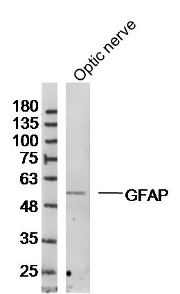 Sample:Optic nerve (Rat)cell Lysate at 40 ug Primary: Anti-GFAP(bs-0199R)at 1/300 dilution Secondary: IRDye800CW Goat Anti-RabbitIgG at 1/20000 dilution Predicted band size: 48 kD Observed band size: 53 kD 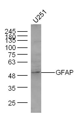 Sample: U251 Cell Lysate at 40 ug Primary: Anti- GFAP (bs-0199R) at 1/300 dilution Secondary: IRDye800CW Goat Anti-Rabbit IgG at 1/10000 dilution Predicted band size: 48 kD Observed band size: 50 kD 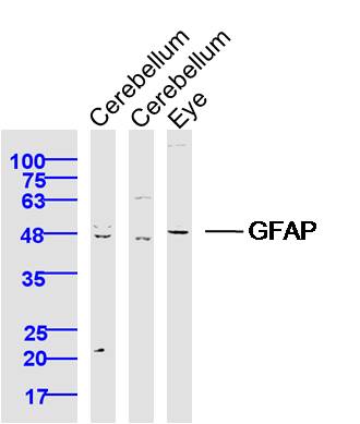 Sample: Cerebellum (Rat) Lysate at 40 ug Cerebellum (Mouse) Lysate at 40 ug Eye (Mouse) Lysate at 40 ug Primary: Anti-GFAP (bs-0199R) at 1/300 dilution Secondary: IRDye800CW Goat Anti-Rabbit IgG at 1/20000 dilution Predicted band size: 48 kD Observed band size: 48 kD 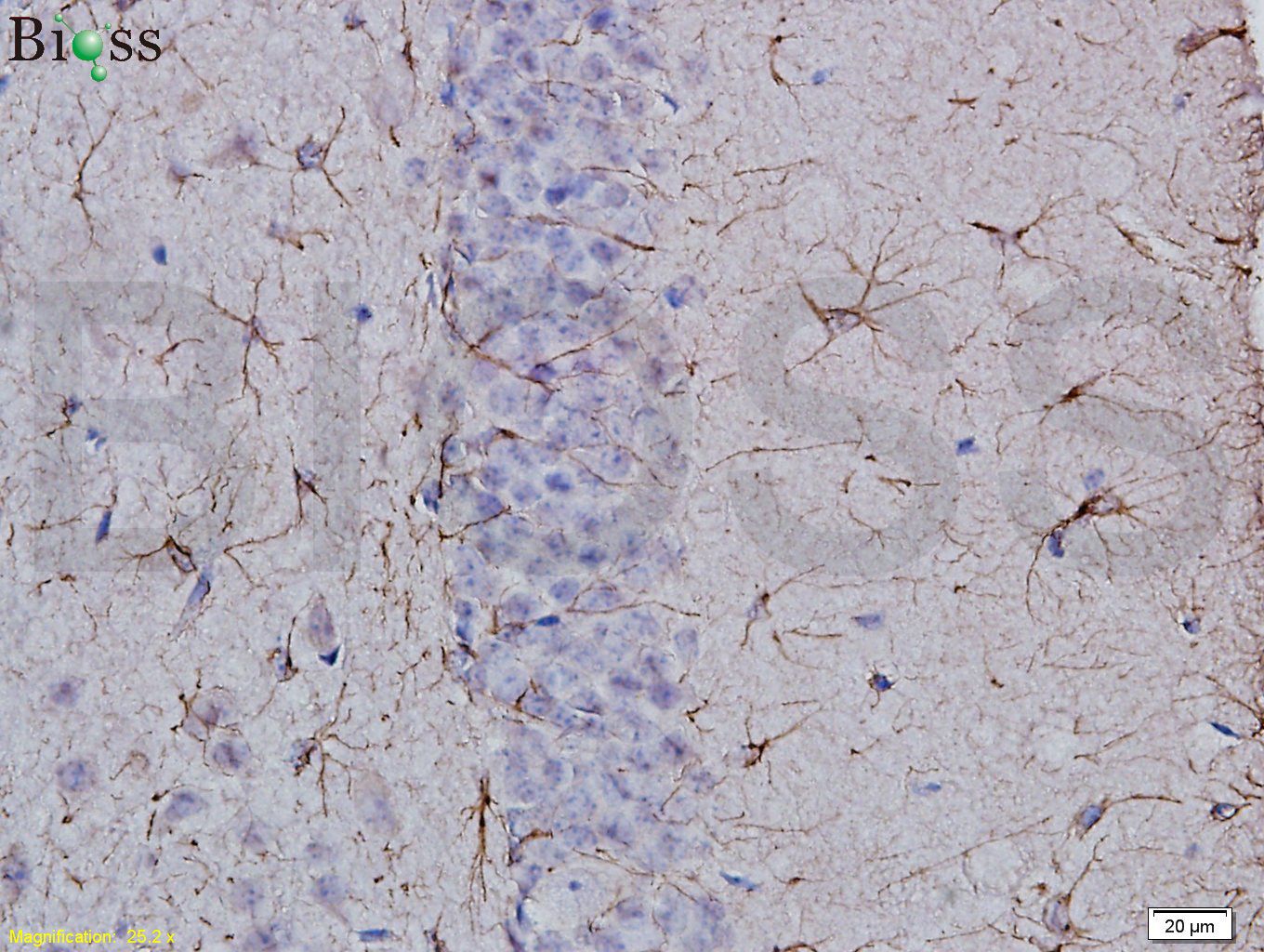 Tissue/cell: rat brain tissue; 4% Paraformaldehyde-fixed and paraffin-embedded; Antigen retrieval: citrate buffer ( 0.01M, pH 6.0 ), Boiling bathing for 15min; Block endogenous peroxidase by 3% Hydrogen peroxide for 30min; Blocking buffer (normal goat serum,C-0005) at 37�� for 20 min; Incubation: Anti-GFAP Polyclonal Antibody, Unconjugated(bs-0199R) 1:400, overnight at 4��C, followed by conjugation to the secondary antibody(SP-0023) and DAB(C-0010) staining 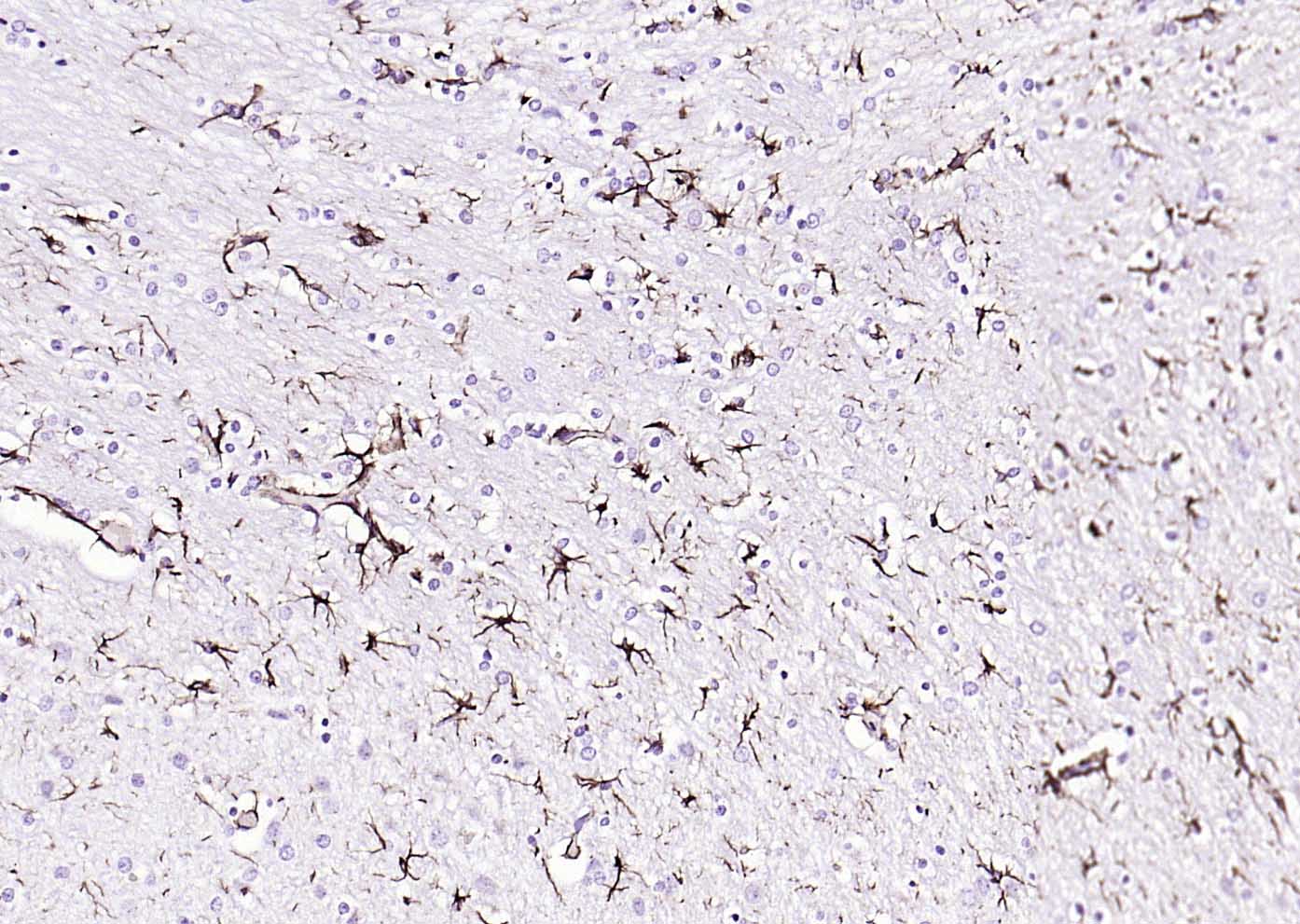 Paraformaldehyde-fixed, paraffin embedded (rat brain); Antigen retrieval by boiling in sodium citrate buffer (pH6.0) for 15min; Block endogenous peroxidase by 3% hydrogen peroxide for 20 minutes; Blocking buffer (normal goat serum) at 37��C for 30min; Antibody incubation with (GFAP) Polyclonal Antibody, Unconjugated (bs-0199R) at 1:200 overnight at 4��C, followed by operating according to SP Kit(Rabbit) (sp-0023) instructionsand DAB staining. 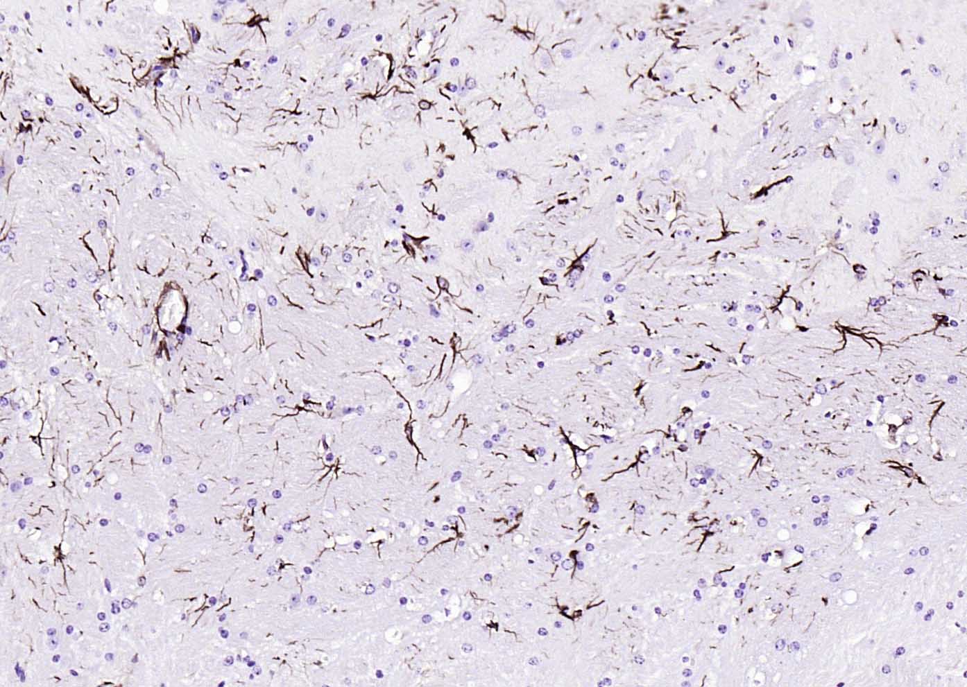 Paraformaldehyde-fixed, paraffin embedded (mouse brain); Antigen retrieval by boiling in sodium citrate buffer (pH6.0) for 15min; Block endogenous peroxidase by 3% hydrogen peroxide for 20 minutes; Blocking buffer (normal goat serum) at 37��C for 30min; Antibody incubation with (GFAP) Polyclonal Antibody, Unconjugated (bs-0199R) at 1:200 overnight at 4��C, followed by operating according to SP Kit(Rabbit) (sp-0023) instructionsand DAB staining. 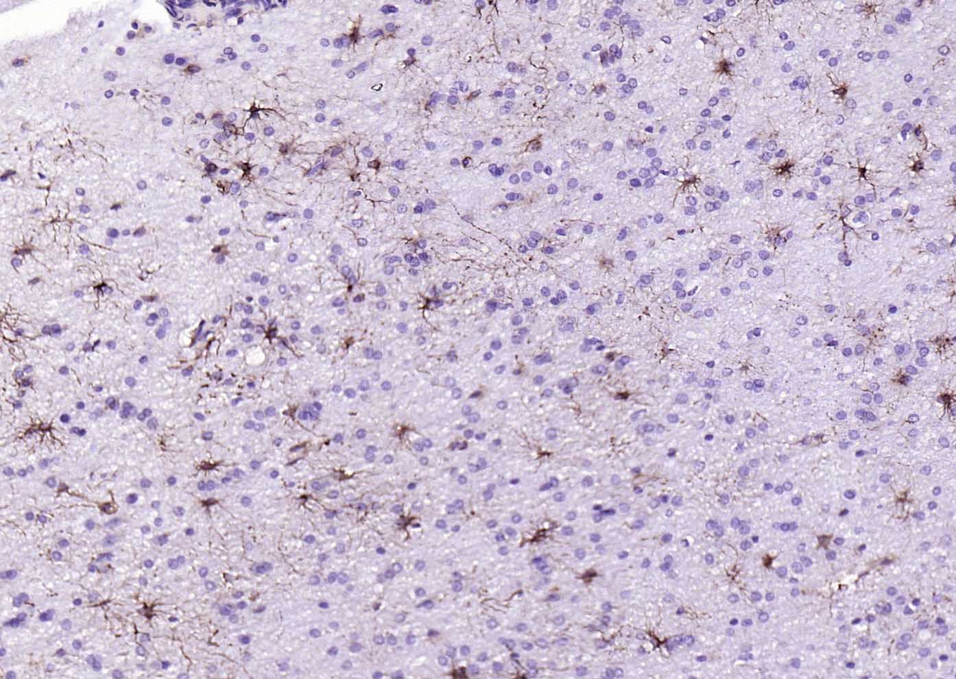 Paraformaldehyde-fixed, paraffin embedded (human glioma); Antigen retrieval by boiling in sodium citrate buffer (pH6.0) for 15min; Block endogenous peroxidase by 3% hydrogen peroxide for 20 minutes; Blocking buffer (normal goat serum) at 37��C for 30min; Antibody incubation with (GFAP) Polyclonal Antibody, Unconjugated (bs-0199R) at 1:200 overnight at 4��C, followed by operating according to SP Kit(Rabbit) (sp-0023) instructionsand DAB staining. 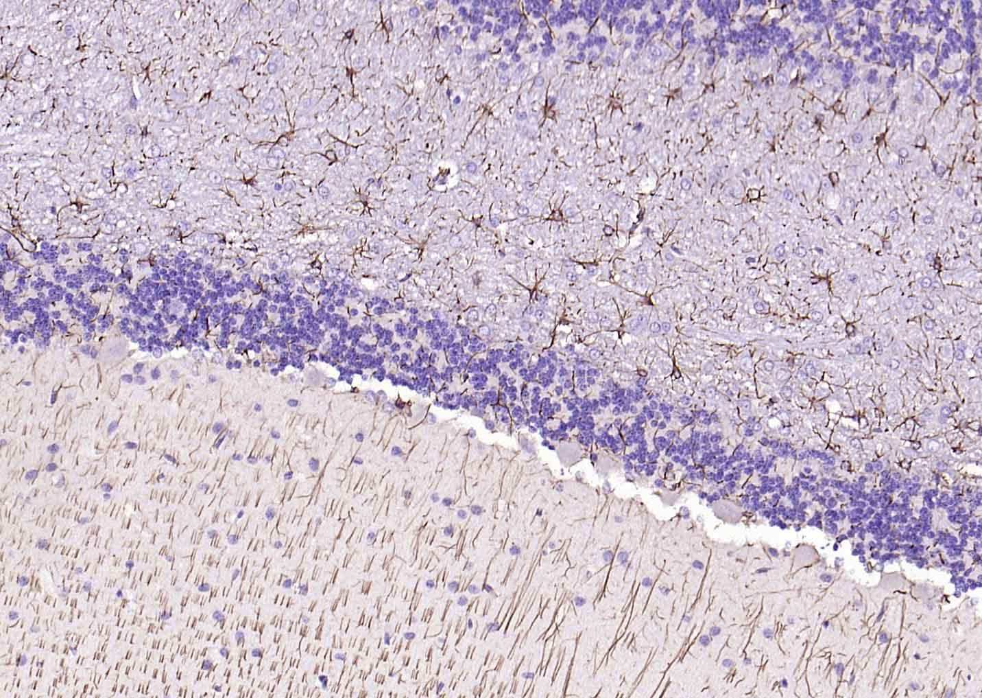 Paraformaldehyde-fixed, paraffin embedded (rat cerebellum); Antigen retrieval by boiling in sodium citrate buffer (pH6.0) for 15min; Block endogenous peroxidase by 3% hydrogen peroxide for 20 minutes; Blocking buffer (normal goat serum) at 37��C for 30min; Antibody incubation with (GFAP) Polyclonal Antibody, Unconjugated (bs-0199R) at 1:200 overnight at 4��C, followed by operating according to SP Kit(Rabbit) (sp-0023) instructionsand DAB staining. 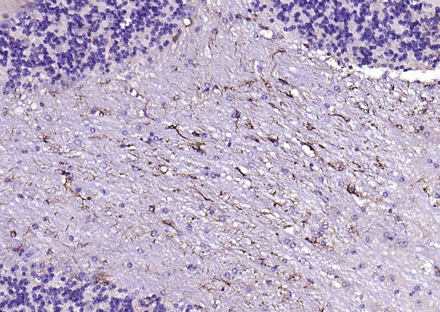 Paraformaldehyde-fixed, paraffin embedded (mouse cerebellum); Antigen retrieval by boiling in sodium citrate buffer (pH6.0) for 15min; Block endogenous peroxidase by 3% hydrogen peroxide for 20 minutes; Blocking buffer (normal goat serum) at 37��C for 30min; Antibody incubation with (GFAP) Polyclonal Antibody, Unconjugated (bs-0199R) at 1:200 overnight at 4��C, followed by operating according to SP Kit(Rabbit) (sp-0023) instructionsand DAB staining. 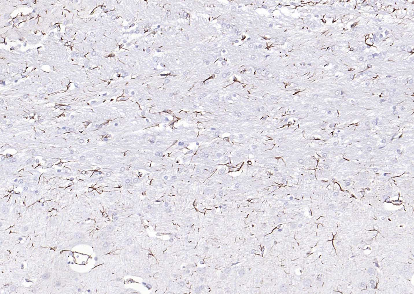 Paraformaldehyde-fixed, paraffin embedded (rat brain); Antigen retrieval by boiling in sodium citrate buffer (pH6.0) for 15min; Block endogenous peroxidase by 3% hydrogen peroxide for 20 minutes; Blocking buffer (normal goat serum) at 37��C for 30min; Antibody incubation with (GFAP) Polyclonal Antibody, Unconjugated (bs-0199R) at 1:200 overnight at 4��C, followed by operating according to SP Kit(Rabbit) (sp-0023) instructionsand DAB staining. 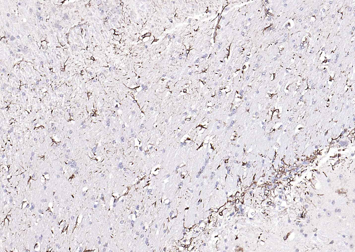 Paraformaldehyde-fixed, paraffin embedded (mouse brain); Antigen retrieval by boiling in sodium citrate buffer (pH6.0) for 15min; Block endogenous peroxidase by 3% hydrogen peroxide for 20 minutes; Blocking buffer (normal goat serum) at 37��C for 30min; Antibody incubation with (GFAP) Polyclonal Antibody, Unconjugated (bs-0199R) at 1:200 overnight at 4��C, followed by operating according to SP Kit(Rabbit) (sp-0023) instructionsand DAB staining. 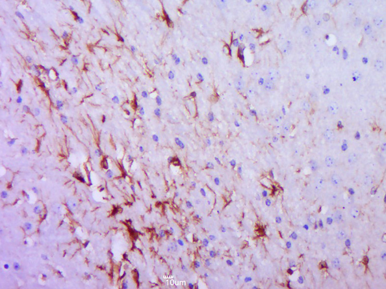 Paraformaldehyde-fixed, paraffin embedded (Mouse brain); Antigen retrieval by boiling in sodium citrate buffer (pH6.0) for 15min; Block endogenous peroxidase by 3% hydrogen peroxide for 20 minutes; Blocking buffer (normal goat serum) at 37��C for 30min; Antibody incubation with (GFAP) Polyclonal Antibody, Unconjugated (bs-0199R) at 1:400 overnight at 4��C, followed by operating according to SP Kit(Rabbit) (sp-0023) instructionsand DAB staining. 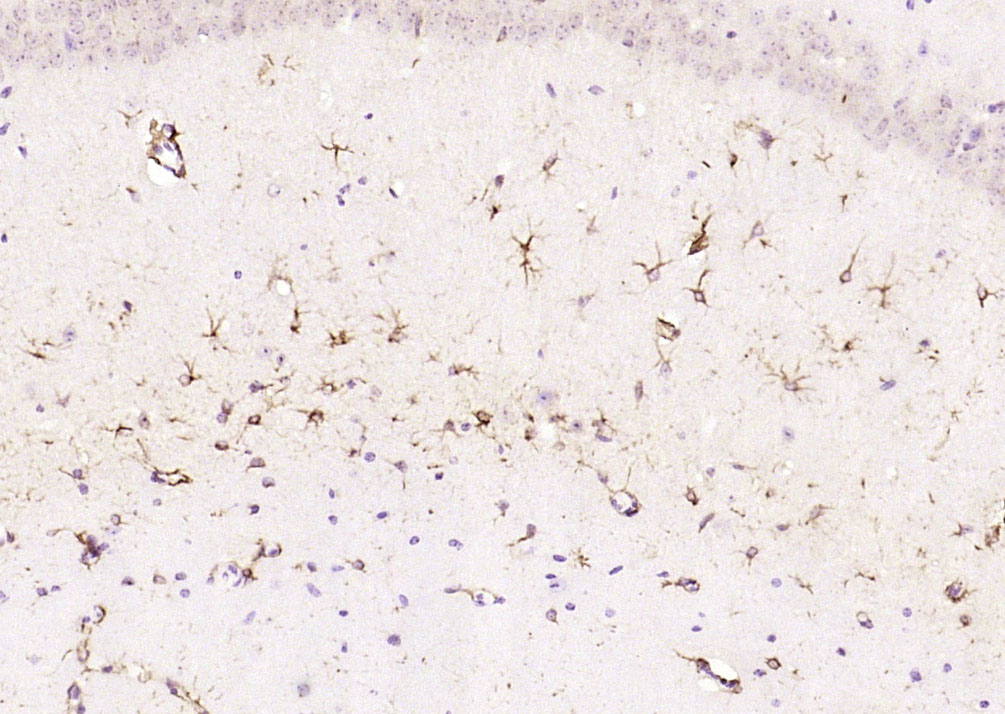 Paraformaldehyde-fixed, paraffin embedded (mouse brain); Antigen retrieval by boiling in sodium citrate buffer (pH6.0) for 15min; Block endogenous peroxidase by 3% hydrogen peroxide for 20 minutes; Blocking buffer (normal goat serum) at 37��C for 30min; Antibody incubation with (GFAP) Polyclonal Antibody, Unconjugated (bs-0199R) at 1:200 overnight at 4��C, followed by operating according to SP Kit(Rabbit) (sp-0023) instructionsand DAB staining. 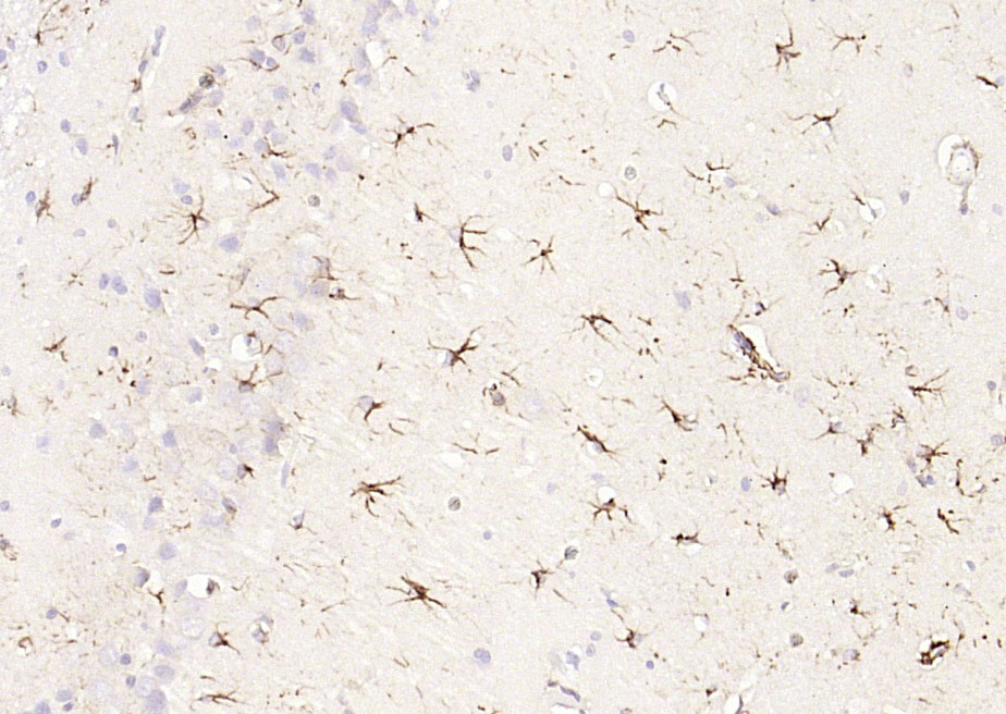 Paraformaldehyde-fixed, paraffin embedded (rat brain); Antigen retrieval by boiling in sodium citrate buffer (pH6.0) for 15min; Block endogenous peroxidase by 3% hydrogen peroxide for 20 minutes; Blocking buffer (normal goat serum) at 37��C for 30min; Antibody incubation with (GFAP) Polyclonal Antibody, Unconjugated (bs-0199R) at 1:200 overnight at 4��C, followed by operating according to SP Kit(Rabbit) (sp-0023) instructionsand DAB staining. 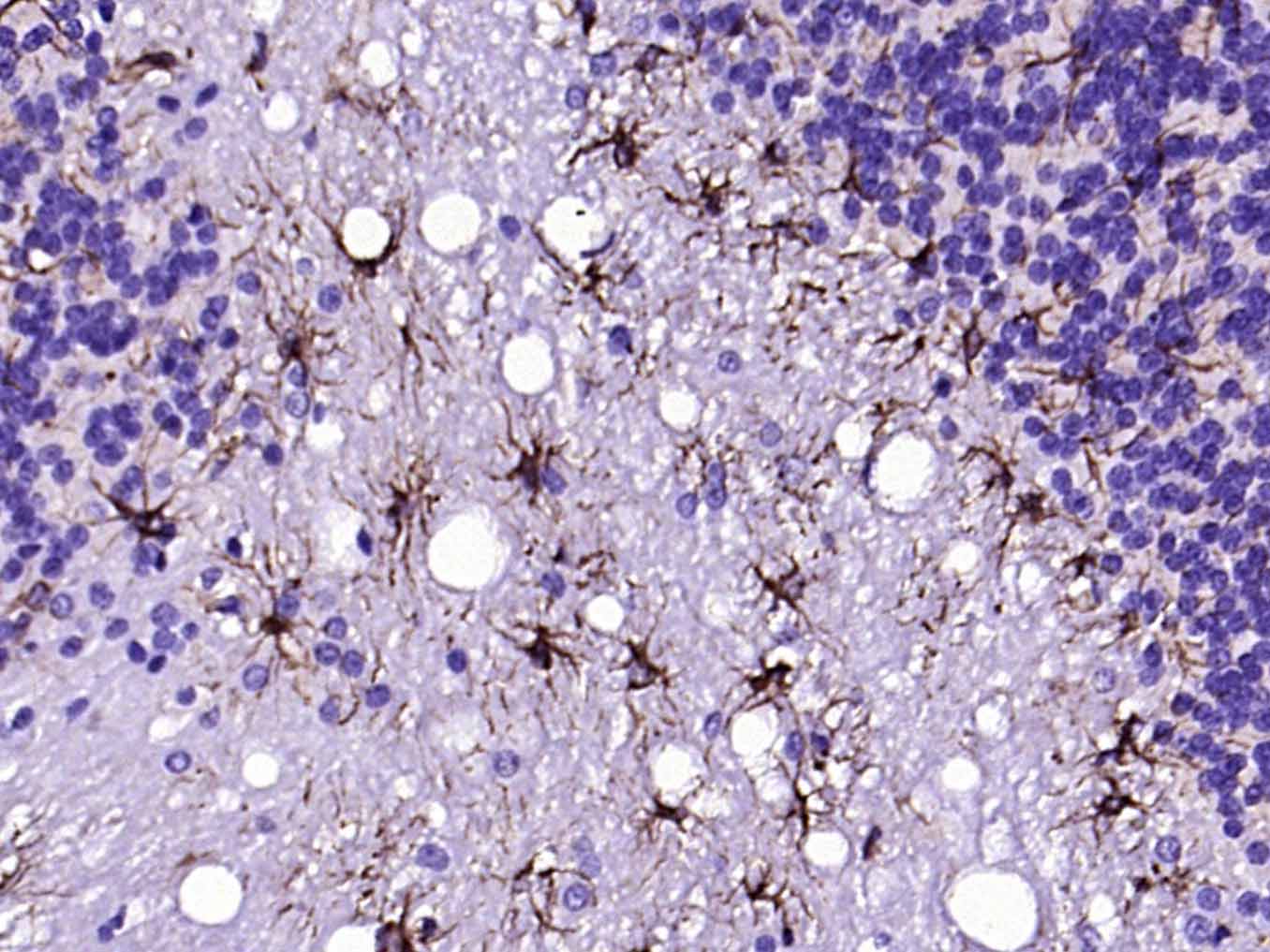 Paraformaldehyde-fixed, paraffin embedded (rat cerebellum); Antigen retrieval by boiling in sodium citrate buffer (pH6.0) for 15min; Block endogenous peroxidase by 3% hydrogen peroxide for 20 minutes; Blocking buffer (normal goat serum) at 37��C for 30min; Antibody incubation with (GFAP) Polyclonal Antibody, Unconjugated (bs-0199R) at 1:200 overnight at 4��C, followed by operating according to SP Kit(Rabbit) (sp-0023) instructionsand DAB staining. 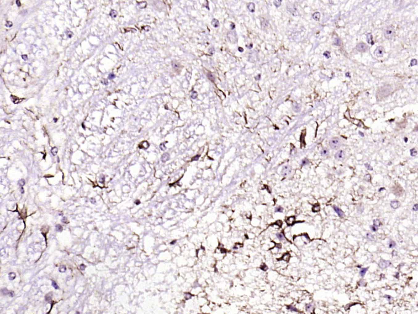 Paraformaldehyde-fixed, paraffin embedded (mouse cerebellum); Antigen retrieval by boiling in sodium citrate buffer (pH6.0) for 15min; Block endogenous peroxidase by 3% hydrogen peroxide for 20 minutes; Blocking buffer (normal goat serum) at 37��C for 30min; Antibody incubation with (GFAP) Polyclonal Antibody, Unconjugated (bs-0199R) at 1:200 overnight at 4��C, followed by operating according to SP Kit(Rabbit) (sp-0023) instructionsand DAB staining. 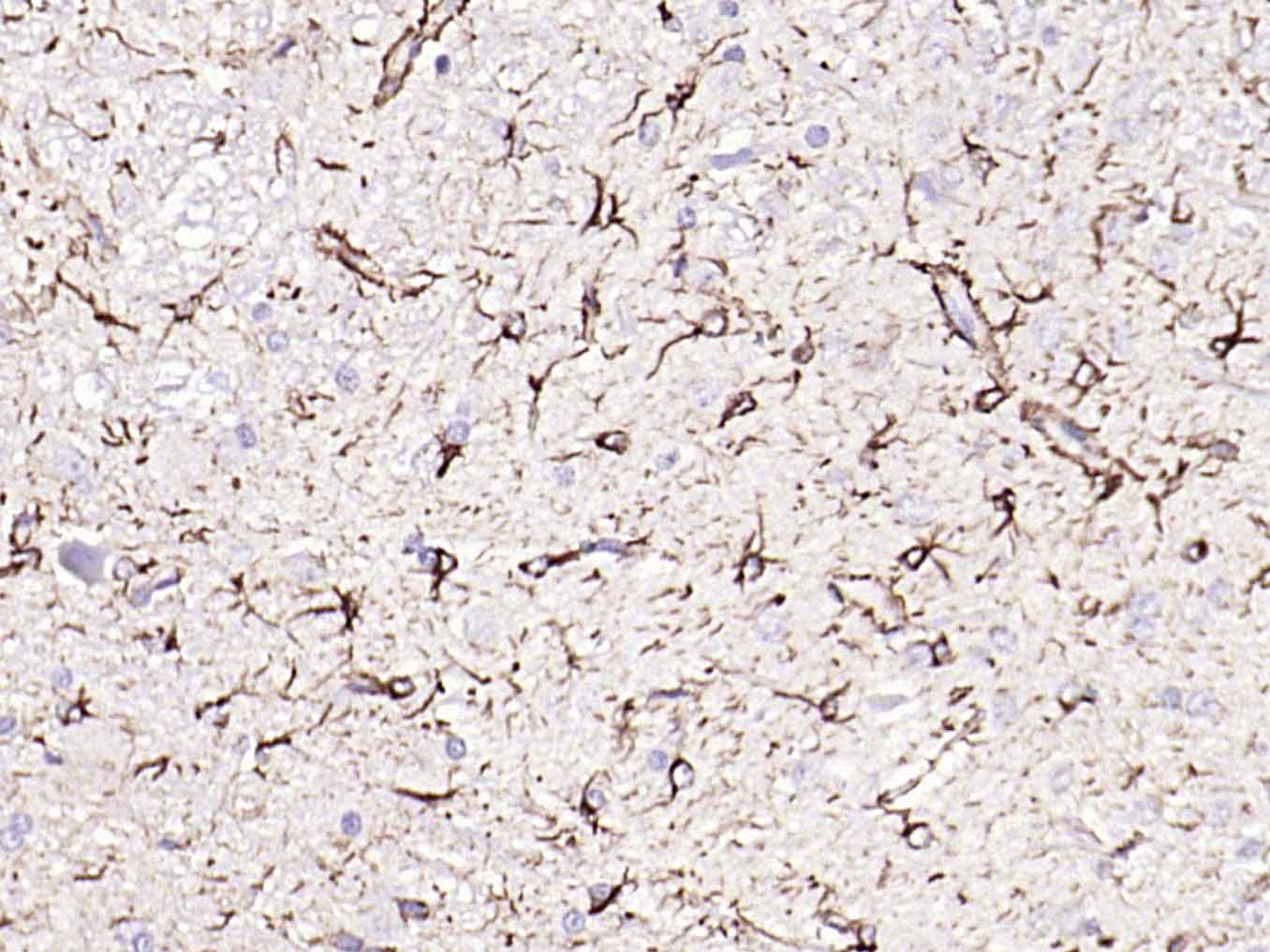 Paraformaldehyde-fixed, paraffin embedded (rat cerebellum); Antigen retrieval by boiling in sodium citrate buffer (pH6.0) for 15min; Block endogenous peroxidase by 3% hydrogen peroxide for 20 minutes; Blocking buffer (normal goat serum) at 37��C for 30min; Antibody incubation with (GFAP) Polyclonal Antibody, Unconjugated (bs-0199R) at 1:200 overnight at 4��C, followed by operating according to SP Kit(Rabbit) (sp-0023) instructionsand DAB staining. 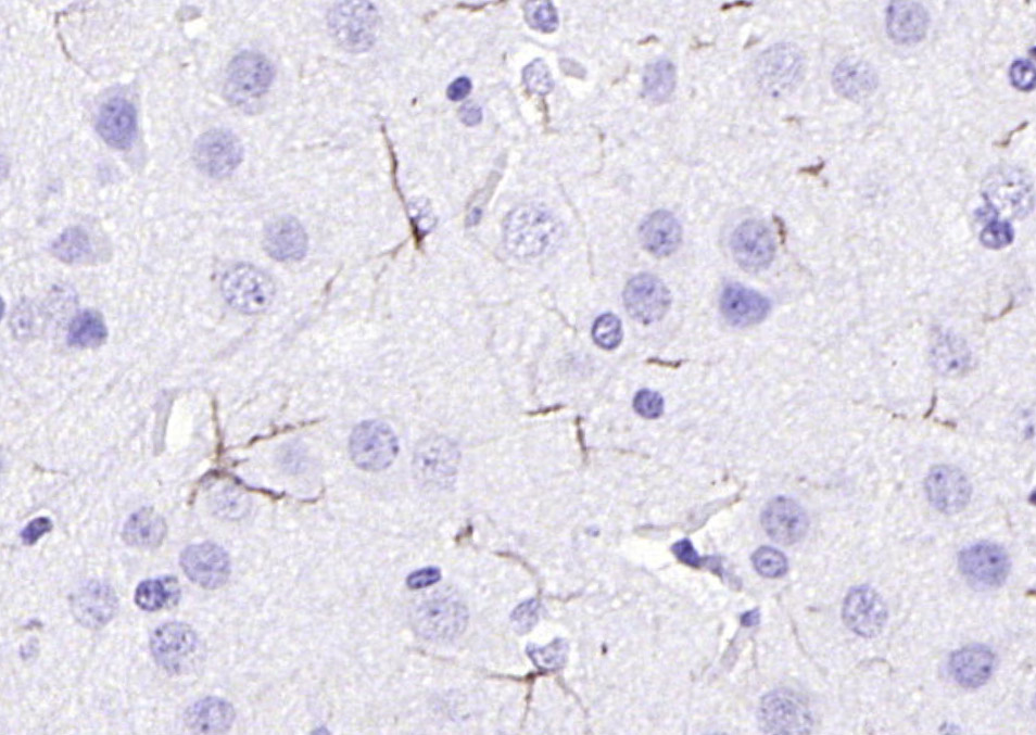 Paraformaldehyde-fixed, paraffin embedded (rat brain); Antigen retrieval by boiling in sodium citrate buffer (pH6.0) for 15min; Block endogenous peroxidase by 3% hydrogen peroxide for 20 minutes; Blocking buffer (normal goat serum) at 37��C for 30min; Antibody incubation with (GFAP) Polyclonal Antibody, Unconjugated (bs-0199R) at 1:200 overnight at 4��C, followed by operating according to SP Kit(Rabbit) (sp-0023) instructionsand DAB staining. 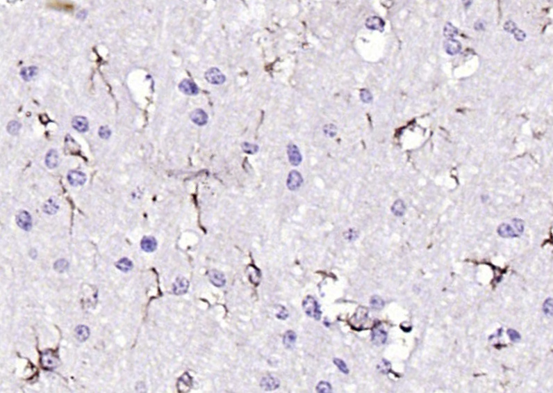 Paraformaldehyde-fixed, paraffin embedded (mouse brain); Antigen retrieval by boiling in sodium citrate buffer (pH6.0) for 15min; Block endogenous peroxidase by 3% hydrogen peroxide for 20 minutes; Blocking buffer (normal goat serum) at 37��C for 30min; Antibody incubation with (GFAP) Polyclonal Antibody, Unconjugated (bs-0199R) at 1:200 overnight at 4��C, followed by operating according to SP Kit(Rabbit) (sp-0023) instructionsand DAB staining. 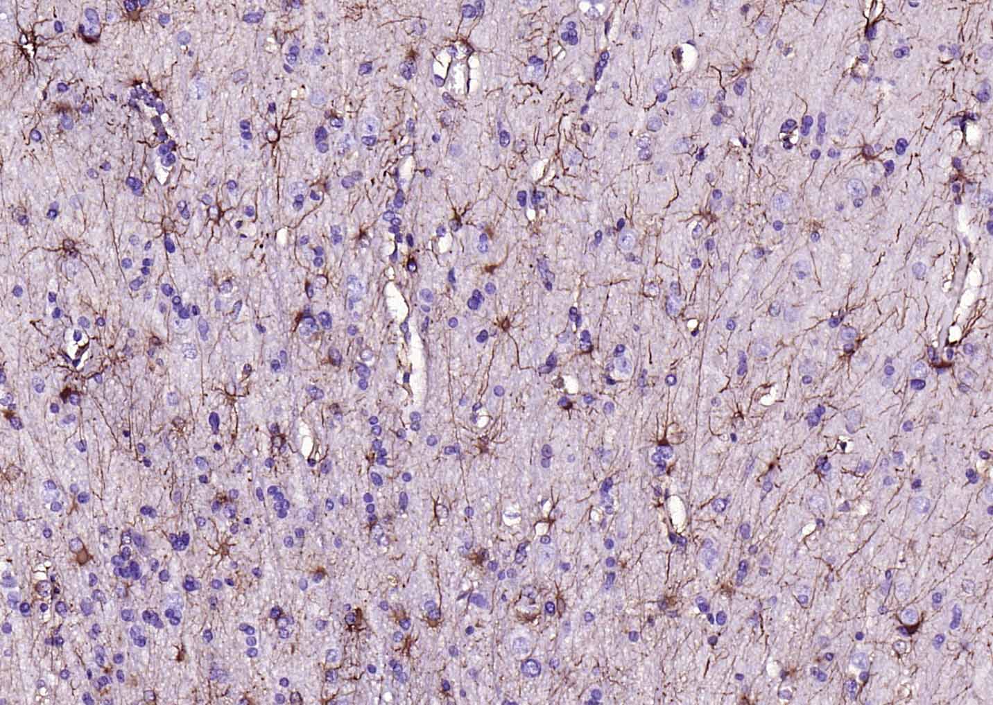 Paraformaldehyde-fixed, paraffin embedded (human brain glioma); Antigen retrieval by boiling in sodium citrate buffer (pH6.0) for 15min; Block endogenous peroxidase by 3% hydrogen peroxide for 20 minutes; Blocking buffer (normal goat serum) at 37��C for 30min; Antibody incubation with (GFAP) Polyclonal Antibody, Unconjugated (bs-0199R) at 1:200 overnight at 4��C, followed by operating according to SP Kit(Rabbit) (sp-0023) instructionsand DAB staining. 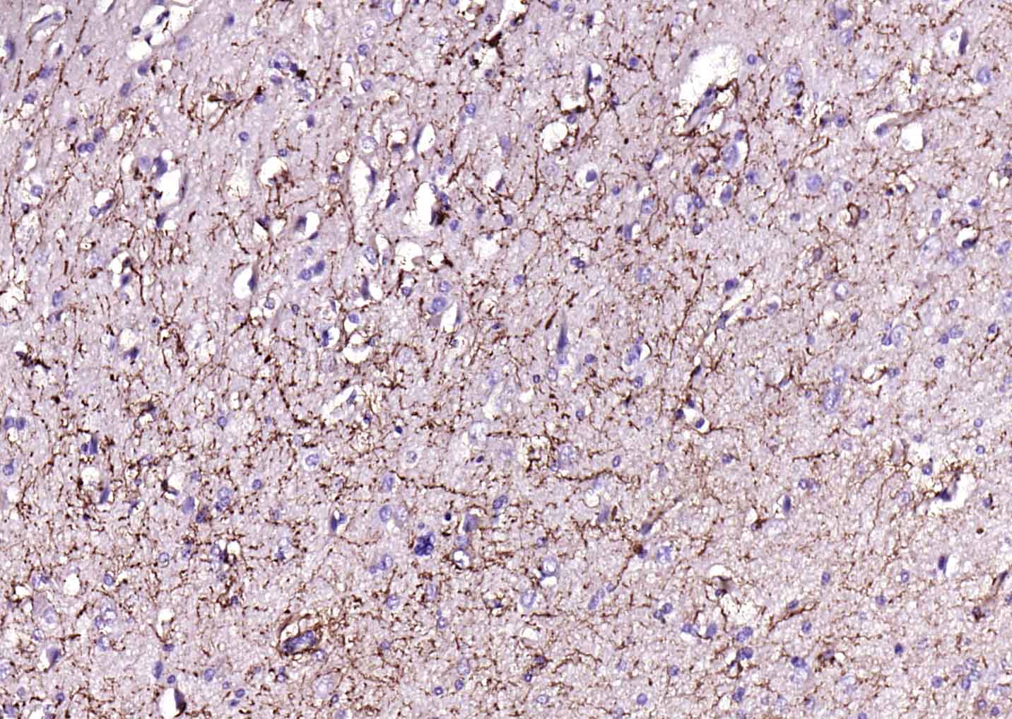 Paraformaldehyde-fixed, paraffin embedded (human brain ); Antigen retrieval by boiling in sodium citrate buffer (pH6.0) for 15min; Block endogenous peroxidase by 3% hydrogen peroxide for 20 minutes; Blocking buffer (normal goat serum) at 37��C for 30min; Antibody incubation with (GFAP) Polyclonal Antibody, Unconjugated (bs-0199R) at 1:200 overnight at 4��C, followed by operating according to SP Kit(Rabbit) (sp-0023) instructionsand DAB staining. 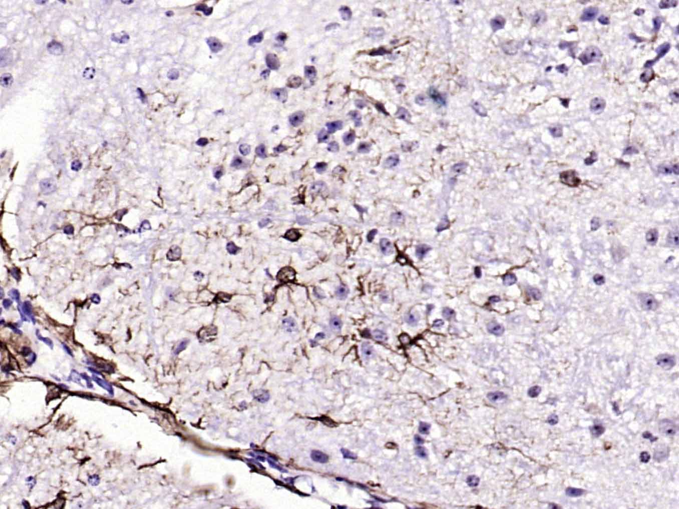 Paraformaldehyde-fixed, paraffin embedded (mouse cerebellum); Antigen retrieval by boiling in sodium citrate buffer (pH6.0) for 15min; Block endogenous peroxidase by 3% hydrogen peroxide for 20 minutes; Blocking buffer (normal goat serum) at 37��C for 30min; Antibody incubation with (GFAP) Polyclonal Antibody, Unconjugated (bs-0199R) at 1:200 overnight at 4��C, followed by operating according to SP Kit(Rabbit) (sp-0023) instructionsand DAB staining. 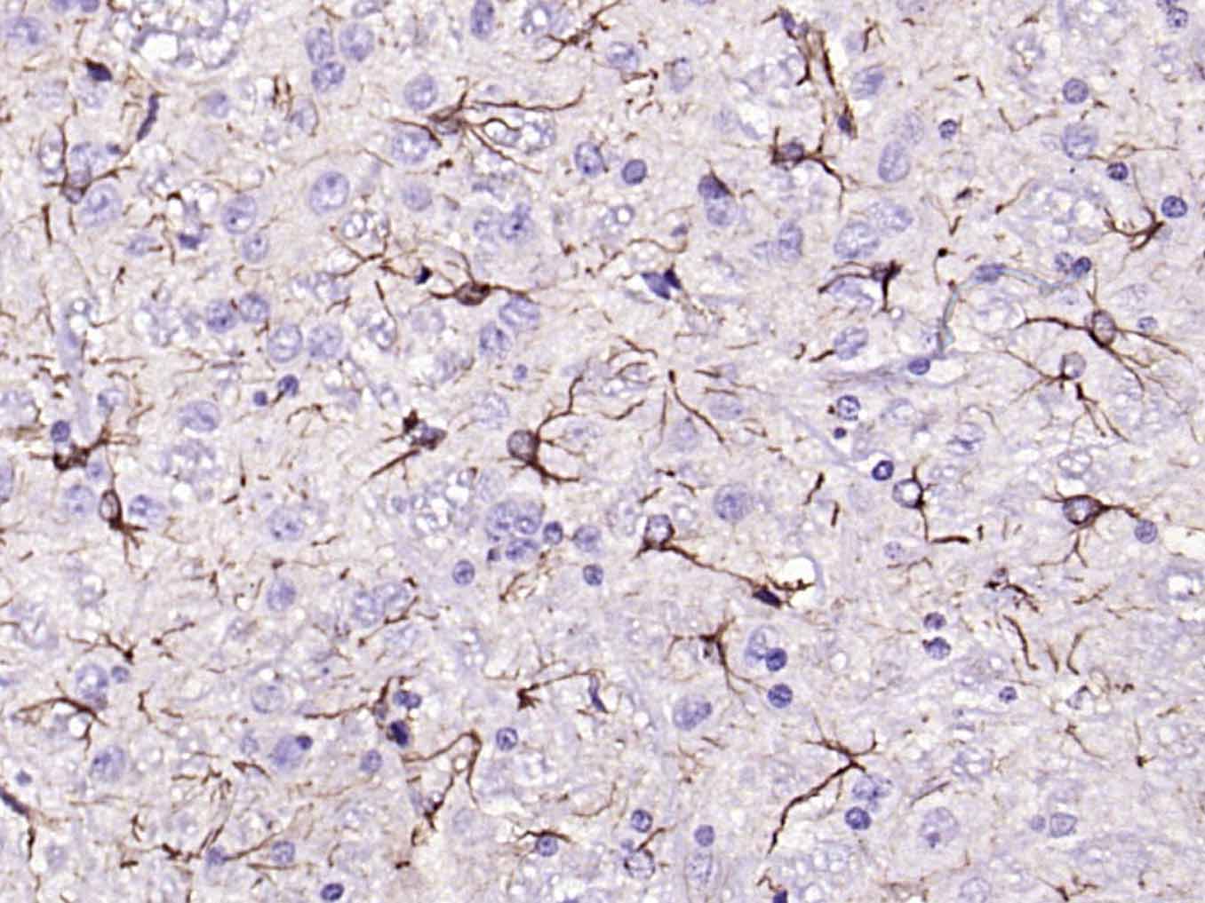 Paraformaldehyde-fixed, paraffin embedded (rat cerebellum); Antigen retrieval by boiling in sodium citrate buffer (pH6.0) for 15min; Block endogenous peroxidase by 3% hydrogen peroxide for 20 minutes; Blocking buffer (normal goat serum) at 37��C for 30min; Antibody incubation with (GFAP) Polyclonal Antibody, Unconjugated (bs-0199R) at 1:200 overnight at 4��C, followed by operating according to SP Kit(Rabbit) (sp-0023) instructionsand DAB staining. 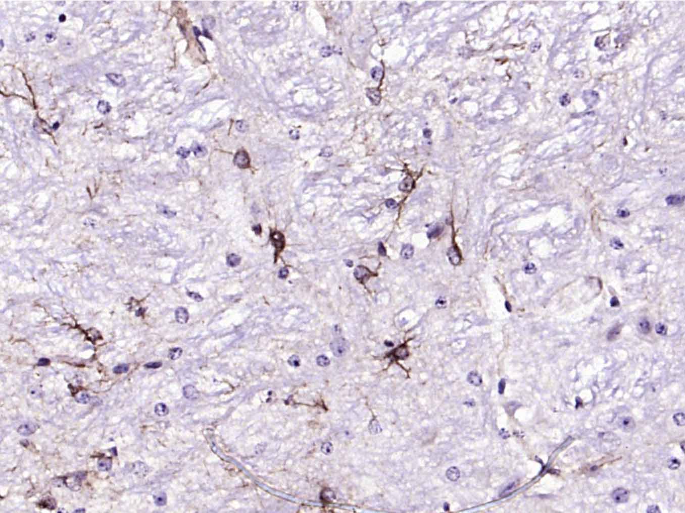 Paraformaldehyde-fixed, paraffin embedded (mouse cerebellum); Antigen retrieval by boiling in sodium citrate buffer (pH6.0) for 15min; Block endogenous peroxidase by 3% hydrogen peroxide for 20 minutes; Blocking buffer (normal goat serum) at 37��C for 30min; Antibody incubation with (GFAP) Polyclonal Antibody, Unconjugated (bs-0199R) at 1:200 overnight at 4��C, followed by operating according to SP Kit(Rabbit) (sp-0023) instructionsand DAB staining. 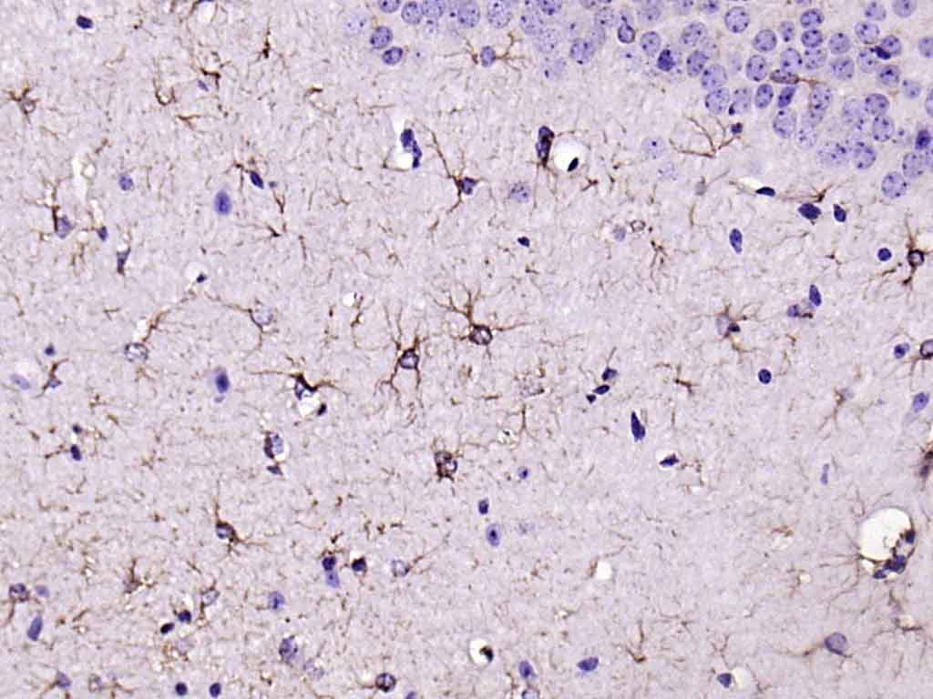 Paraformaldehyde-fixed, paraffin embedded (mouse brain); Antigen retrieval by boiling in sodium citrate buffer (pH6.0) for 15min; Block endogenous peroxidase by 3% hydrogen peroxide for 20 minutes; Blocking buffer (normal goat serum) at 37��C for 30min; Antibody incubation with (GFAP) Polyclonal Antibody, Unconjugated (bs-0199R) at 1:200 overnight at 4��C, followed by operating according to SP Kit(Rabbit) (sp-0023) instructionsand DAB staining. 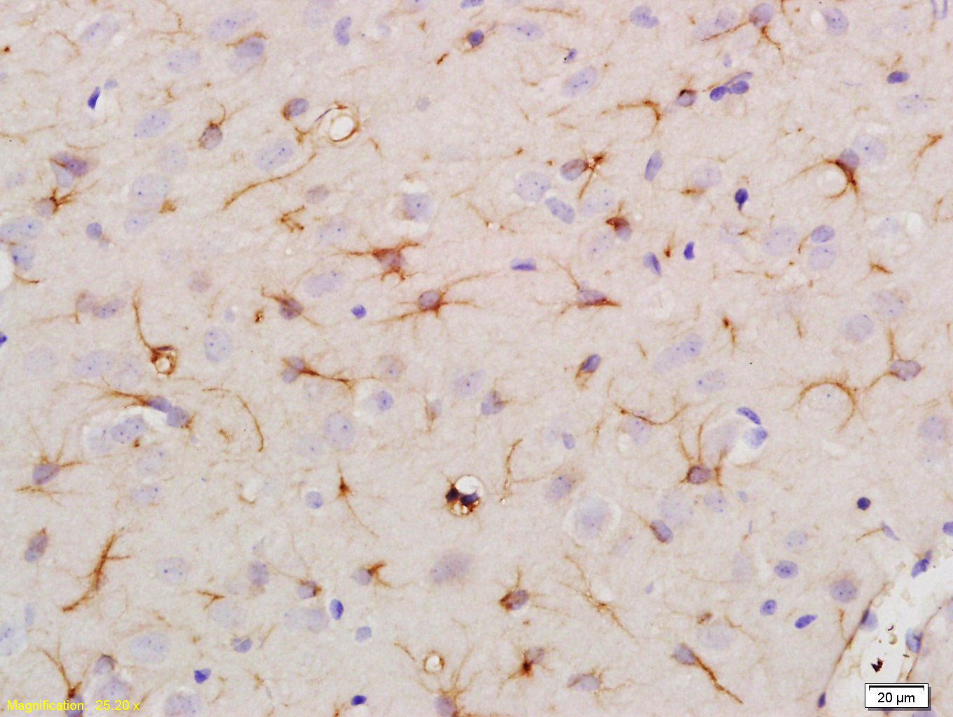 Tissue/cell: rat brain tissue; 4% Paraformaldehyde-fixed and paraffin-embedded; Antigen retrieval: citrate buffer ( 0.01M, pH 6.0 ), Boiling bathing for 15min; Block endogenous peroxidase by 3% Hydrogen peroxide for 30min; Blocking buffer (normal goat serum,C-0005) at 37�� for 20 min; Incubation: Anti-GFAP Polyclonal Antibody, Unconjugated(bs-0199R) 1:500, overnight at 4��C, followed by conjugation to the secondary antibody(SP-0023) and DAB(C-0010) staining 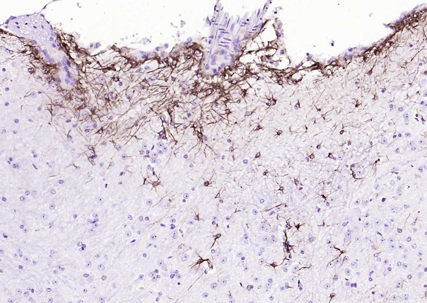 Paraformaldehyde-fixed, paraffin embedded (mouse cerebellum); Antigen retrieval by boiling in sodium citrate buffer (pH6.0) for 15min; Block endogenous peroxidase by 3% hydrogen peroxide for 20 minutes; Blocking buffer (normal goat serum) at 37��C for 30min; Antibody incubation with (GFAP) Polyclonal Antibody, Unconjugated (bs-0199R) at 1:200 overnight at 4��C, followed by operating according to SP Kit(Rabbit) (sp-0023) instructionsand DAB staining. 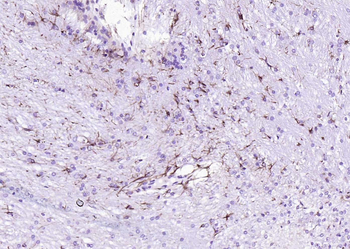 Paraformaldehyde-fixed, paraffin embedded (rat cerebellum); Antigen retrieval by boiling in sodium citrate buffer (pH6.0) for 15min; Block endogenous peroxidase by 3% hydrogen peroxide for 20 minutes; Blocking buffer (normal goat serum) at 37��C for 30min; Antibody incubation with (GFAP) Polyclonal Antibody, Unconjugated (bs-0199R) at 1:200 overnight at 4��C, followed by operating according to SP Kit(Rabbit) (sp-0023) instructionsand DAB staining. 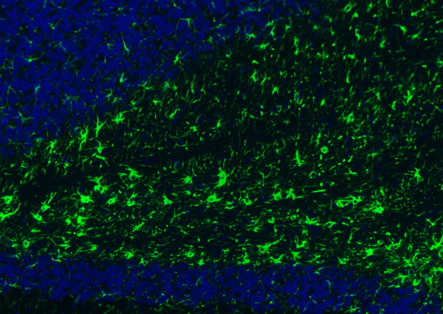 Paraformaldehyde-fixed, paraffin embedded (rat cerebellum); Antigen retrieval by boiling in sodium citrate buffer (pH6.0) for 15min; Blocking buffer (normal goat serum) at 37��C for 30min; Antibody incubation with (GFAP) Polyclonal Antibody, Unconjugated (bs-0199R) at 1:300 overnight at 4��C, followed by a conjugated Goat Anti-Rabbit IgG antibody (YF488) for 90 minutes, and DAPI for nuclei staining. 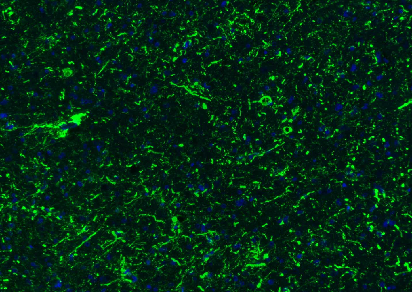 Paraformaldehyde-fixed, paraffin embedded (human brain); Antigen retrieval by boiling in sodium citrate buffer (pH6.0) for 15min; Blocking buffer (normal goat serum) at 37��C for 30min; Antibody incubation with (GFAP) Polyclonal Antibody, Unconjugated (bs-0199R) at 1:500 overnight at 4��C, followed by a conjugated Goat Anti-Rabbit IgG antibody (YF488) for 90 minutes, and DAPI for nuclei staining. 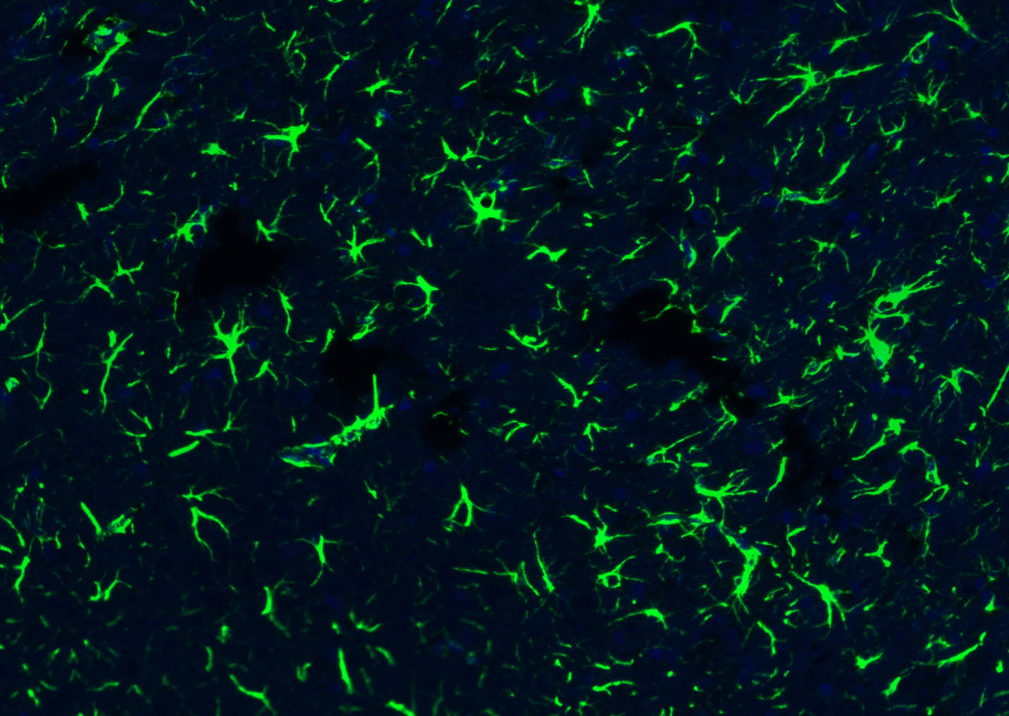 Paraformaldehyde-fixed, paraffin embedded (rat brain); Antigen retrieval by boiling in sodium citrate buffer (pH6.0) for 15min; Blocking buffer (normal goat serum) at 37��C for 30min; Antibody incubation with (GFAP) Polyclonal Antibody, Unconjugated (bs-0199R) at 1:500 overnight at 4��C, followed by a conjugated Goat Anti-Rabbit IgG antibody (YF488) for 90 minutes, and DAPI for nuclei staining. 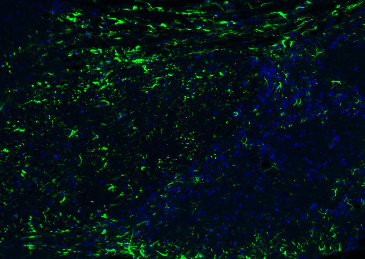 Paraformaldehyde-fixed, paraffin embedded (mouse cerebellum); Antigen retrieval by boiling in sodium citrate buffer (pH6.0) for 15min; Blocking buffer (normal goat serum) at 37��C for 30min; Antibody incubation with (GFAP) Polyclonal Antibody, Unconjugated (bs-0199R) at 1:500 overnight at 4��C, followed by a conjugated Goat Anti-Rabbit IgG antibody (YF488) for 90 minutes, and DAPI for nuclei staining. 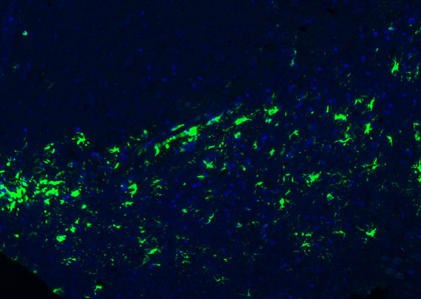 Paraformaldehyde-fixed, paraffin embedded (mouse brain); Antigen retrieval by boiling in sodium citrate buffer (pH6.0) for 15min; Blocking buffer (normal goat serum) at 37��C for 30min; Antibody incubation with (GFAP) Polyclonal Antibody, Unconjugated (bs-0199R) at 1:500 overnight at 4��C, followed by a conjugated Goat Anti-Rabbit IgG antibody (YF488) for 90 minutes, and DAPI for nuclei staining. 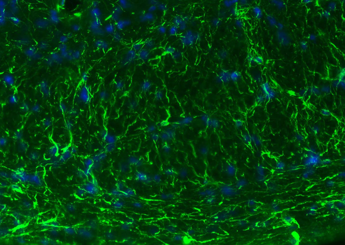 Paraformaldehyde-fixed, paraffin embedded (mouse cerebellum); Antigen retrieval by boiling in sodium citrate buffer (pH6.0) for 15min; Blocking buffer (normal goat serum) at 37��C for 30min; Antibody incubation with (GFAP) Polyclonal Antibody, Unconjugated (bs-0199R) at 1:200 overnight at 4��C, followed by a conjugated Goat Anti-Rabbit IgG antibody (bs-0295G-FITC) for 90 minutes, and DAPI for nuclei staining. 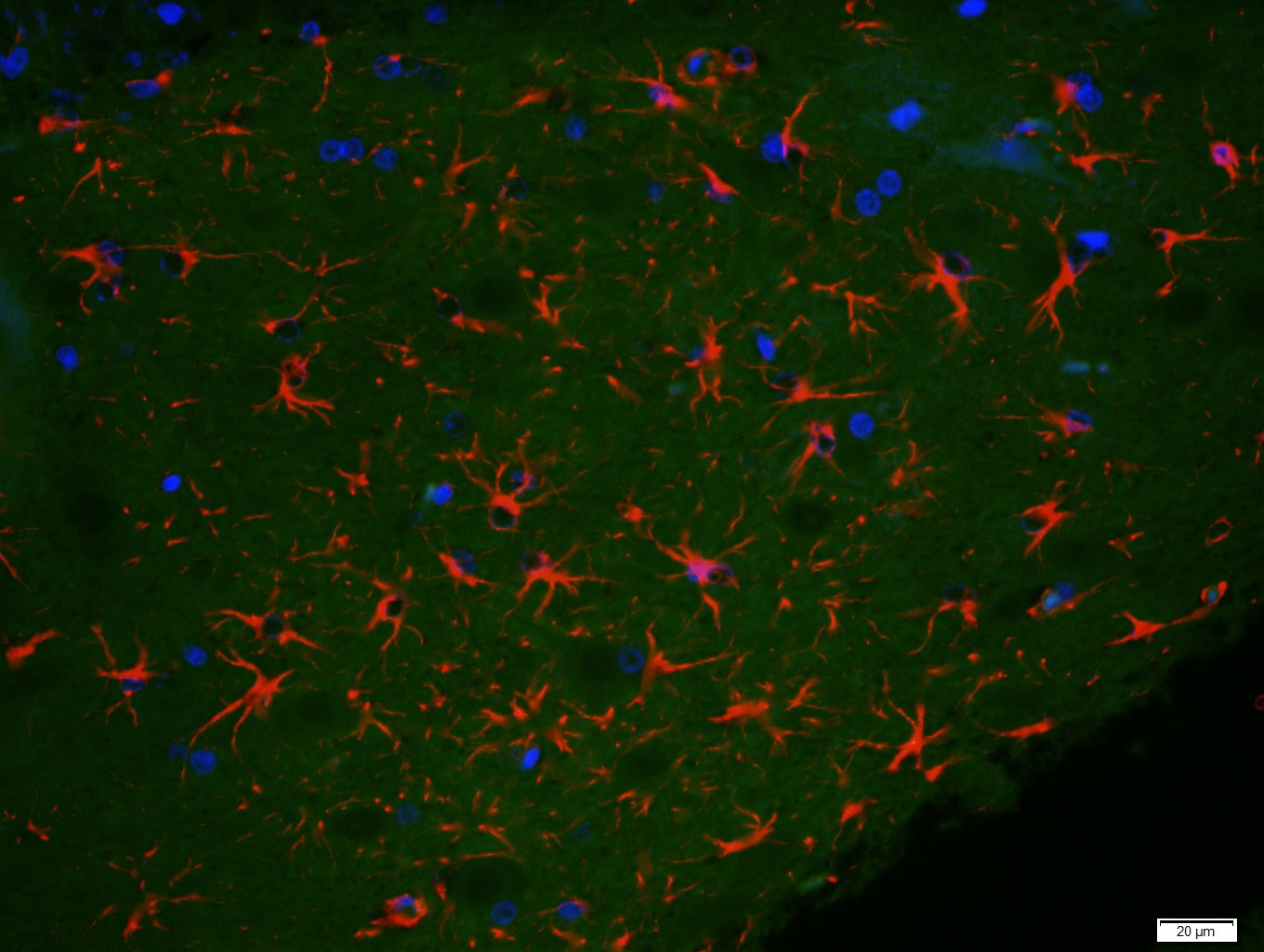 Paraformaldehyde-fixed, paraffin embedded (rat brain); Antigen retrieval by boiling in sodium citrate buffer (pH6) for 15min; Block endogenous peroxidase by 3% hydrogen peroxide for 20 minutes; Blocking buffer (normal goat serum) at 37��C for 30min; Antibody incubation with (GFAP) Polyclonal Antibody, Unconjugated (bs-0199R) at 1:200 overnight at 4��C, followed by a conjugated secondary (bs-0295G-Cy3) at [1:500] for 90 minutes and DAPI staining of the nuclei. 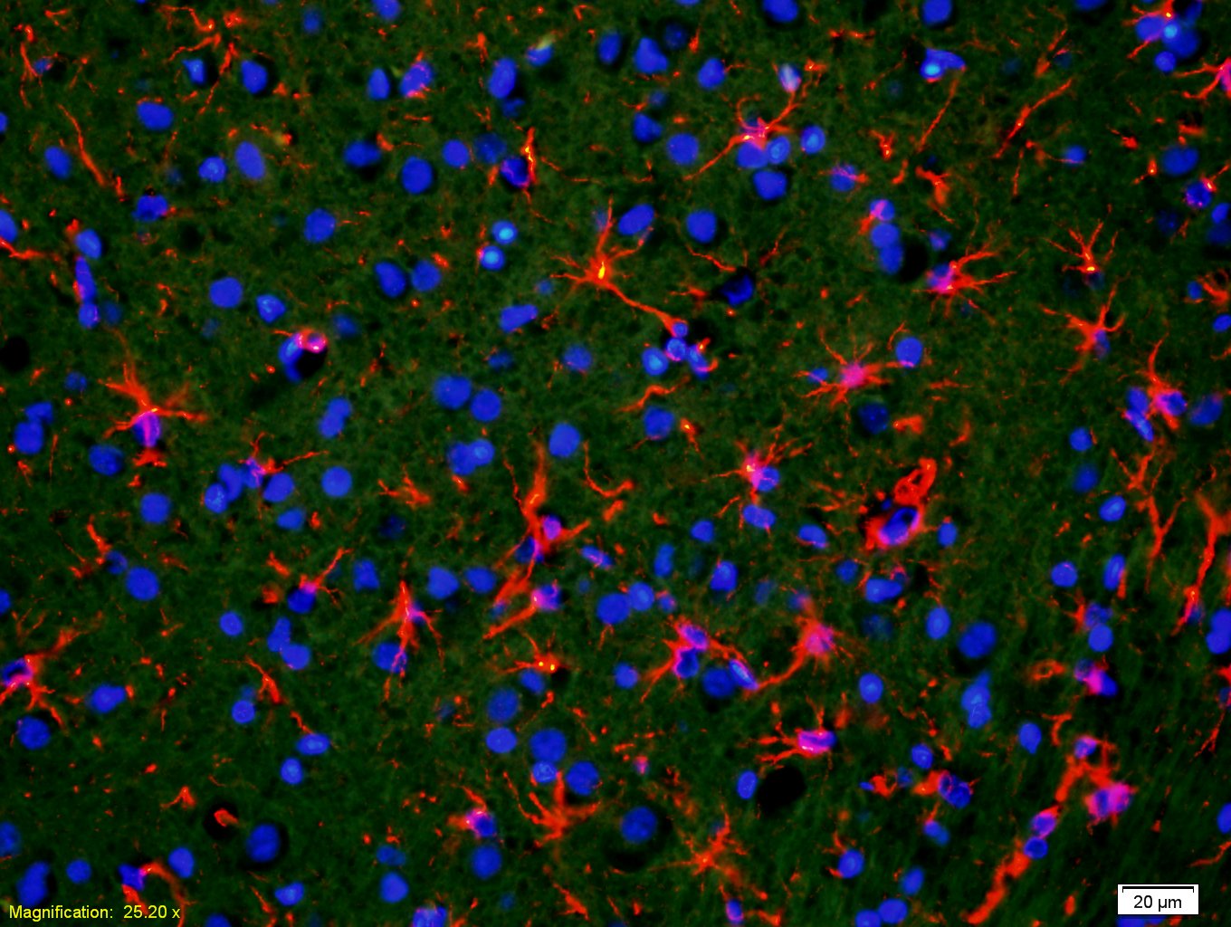 Tissue/cell: rat brain tissue;4% Paraformaldehyde-fixed and paraffin-embedded; Antigen retrieval: citrate buffer ( 0.01M, pH 6.0 ), Boiling bathing for 15min; Blocking buffer (normal goat serum,C-0005) at 37�� for 20 min; Incubation: Anti-GFAP Polyclonal Antibody, Unconjugated(bs-0199R) 1:400, overnight at 4��C; The secondary antibody was Goat Anti-Rabbit IgG, Cy3 conjugated(bs-0295G-Cy3)used at 1:200 dilution for 40 minutes at 37��C. DAPI(5ug/ml,blue,C-0033) was used to stain the cell nuclei 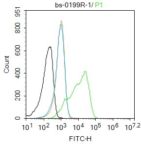 Blank control:U87MG. Primary Antibody (green line): Rabbit Anti-GFAP antibody (bs-0199R) Dilution: 1ug/Test; Secondary Antibody : Goat anti-rabbit IgG-FITC Dilution: 0.5ug/Test. Protocol The cells were fixed with 4% PFA (10min at room temperature)and then permeabilized with 90% ice-cold methanol for 20 min at -20��.The cells were then incubated in 5%BSA to block non-specific protein-protein interactions for 30 min at room temperature .Cells stained with Primary Antibody for 30 min at room temperature. The secondary antibody used for 40 min at room temperature. Acquisition of 20,000 events was performed. 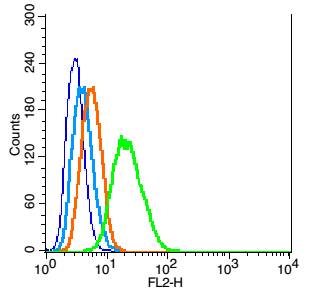 Blank control: RSC96(blue). Primary Antibody:Rabbit Anti- GFAP antibody(bs-0199R), Dilution: 1��g in 100 ��L 1X PBS containing 0.5% BSA; Isotype Control Antibody: Rabbit IgG(orange) ,used under the same conditions ); Secondary Antibody: Goat anti-rabbit IgG-PE(white blue), Dilution: 1:200 in 1 X PBS containing 0.5% BSA. Protocol The cells were fixed with 2% paraformaldehyde (10 min) , then permeabilized with 90% ice-cold methanol for 30 min on ice. Primary antibody (bs-0199R, 1��g /1x10^6 cells) were incubated for 30 min on the ice, followed by 1 X PBS containing 0.5% BSA + 1 0% goat serum (15 min) to block non-specific protein-protein interactions. Then the Goat Anti-rabbit IgG/PE antibody was added into the blocking buffer mentioned above to react with the primary antibody at 1/200 dilution for 30 min on ice. Acquisition of 20,000 events was performed. |


