
上海细胞库
人源细胞系| 稳转细胞系| 基因敲除株| 基因点突变细胞株| 基因过表达细胞株| 重组细胞系| 猪的细胞系| 马细胞系| 兔的细胞系| 犬的细胞系| 山羊的细胞系| 鱼的细胞系| 猴的细胞系| 仓鼠的细胞系| 狗的细胞系| 牛的细胞| 大鼠细胞系| 小鼠细胞系| 其他细胞系|
人源细胞系| 稳转细胞系| 基因敲除株| 基因点突变细胞株| 基因过表达细胞株| 重组细胞系| 猪的细胞系| 马细胞系| 兔的细胞系| 犬的细胞系| 山羊的细胞系| 鱼的细胞系| 猴的细胞系| 仓鼠的细胞系| 狗的细胞系| 牛的细胞| 大鼠细胞系| 小鼠细胞系| 其他细胞系|

| 规格 | 价格 | 库存 |
|---|---|---|
| 50ul | ¥ 1200 | 7 |
| 100ul | ¥ 1900 | 7 |
| 200ul | ¥ 2900 | 6 |
| 产品编号 | Ys-1870R |
| 英文名称 | PEG3 |
| 中文名称 | PEG3蛋白抗体 |
| 别 名 | Paternally-expressed gene 3 protein; Zinc finger and SCAN domain-containing protein 24; Kruppel-type zinc finger protein; paternally expressed 3; Paternally expressed gene 3; PEG3 protein; Pw1; Zn-finger protein Pw1; PEG3_HUMAN; Paternally-expressed gene 3 protein. |
| 抗体来源 | Rabbit |
| 克隆类型 | Polyclonal |
| 交叉反应 | Human, Mouse, Rat, (predicted: Cow, Horse, ) |
| 产品应用 | ELISA=1:5000-10000 IHC-P=1:100-500 IHC-F=1:100-500 IF=1:100-500 (石蜡切片需做抗原修复) not yet tested in other applications. optimal dilutions/concentrations should be determined by the end user. |
| 理论分子量 | 175kDa |
| 细胞定位 | 细胞核 细胞浆 |
| 性 状 | Liquid |
| 浓 度 | 1mg/ml |
| 免 疫 原 | KLH conjugated synthetic peptide derived from human PEG3: 1501-1588/1588 |
| 亚 型 | IgG |
| 纯化方法 | affinity purified by Protein A |
| 缓 冲 液 | 0.01M TBS(pH7.4) with 1% BSA, 0.03% Proclin300 and 50% Glycerol. |
| 保存条件 | Shipped at 4℃. Store at -20 °C for one year. Avoid repeated freeze/thaw cycles. |
| 注意事项 | This product as supplied is intended for research use only, not for use in human, therapeutic or diagnostic applications. |
| PubMed | PubMed |
| 产品介绍 | In human, ZIM2 and PEG3 are treated as two distinct genes though they share multiple 5' exons and a common promoter and both genes are paternally expressed (PMID:15203203). Alternative splicing events connect their shared 5' exons either with the remaining 4 exons unique to ZIM2, or with the remaining 2 exons unique to PEG3. In contrast, in other mammals ZIM2 does not undergo imprinting and, in mouse, cow, and likely other mammals as well, the ZIM2 and PEG3 genes do not share exons. Human PEG3 protein belongs to the Kruppel C2H2-type zinc finger protein family. PEG3 may play a role in cell proliferation and p53-mediated apoptosis. PEG3 has also shown tumor suppressor activity and tumorigenesis in glioma and ovarian cells. Alternative splicing of this PEG3 gene results in multiple transcript variants encoding distinct isoforms. Function: Induces apoptosis in cooperation with SIAH1A. Acts as a mediator between p53/TP53 and BAX in a neuronal death pathway that is activated by DNA damage. Acts synergistically with TRAF2 and inhibits TNF induced apoptosis through activation of NF-kappa-B. Possesses a tumor suppressing activity in glioma cells. Subunit: Homodimer. Interacts with SIAH1A and SIAH2. Interacts with TRAF2. Subcellular Location: Nucleus. Cytoplasm. Tissue Specificity: Brain, glial cells, astrocytes, embryo, placenta, testis, ovary and uterus. In the placenta it is found in the layer of villous cytotrophoblast cells while in the ovary it is found in the cells of the ovarian stroma including the thecal layers around the follicles. Expression is highly repressed in glioma cell lines. Similarity: Belongs to the krueppel C2H2-type zinc-finger protein family. Contains 12 C2H2-type zinc fingers. Contains 1 SCAN box domain. SWISS: Q9GZU2 Gene ID: 18616 PEG3蛋白是一种转录因子。在胰腺、脑组织、胎盘、卵巢、睾丸等组织中表达,PEG3蛋白位于细胞核内。PEG3也是一个凋亡促进因子并可能参与调节肿瘤坏死因子(TNF)信号传导通路。 |
| 产品图片 | Paraformaldehyde-fixed, paraffin embedded (mouse brain); Antigen retrieval by boiling in sodium citrate buffer (pH6.0) for 15min; Block endogenous peroxidase by 3% hydrogen peroxide for 20 minutes; Blocking buffer (normal goat serum) at 37°C for 30min; Antibody incubation with (PEG3) Polyclonal Antibody, Unconjugated (bs-1870R) at 1:200 overnight at 4°C, followed by operating according to SP Kit(Rabbit) (sp-0023) instructionsand DAB staining. Paraformaldehyde-fixed, paraffin embedded (mouse ovary); Antigen retrieval by boiling in sodium citrate buffer (pH6.0) for 15min; Block endogenous peroxidase by 3% hydrogen peroxide for 20 minutes; Blocking buffer (normal goat serum) at 37°C for 30min; Antibody incubation with (PEG3) Polyclonal Antibody, Unconjugated (bs-1870R) at 1:200 overnight at 4°C, followed by operating according to SP Kit(Rabbit) (sp-0023) instructionsand DAB staining. Paraformaldehyde-fixed, paraffin embedded (human brain glioma); Antigen retrieval by boiling in sodium citrate buffer (pH6.0) for 15min; Block endogenous peroxidase by 3% hydrogen peroxide for 20 minutes; Blocking buffer (normal goat serum) at 37°C for 30min; Antibody incubation with (PEG3) Polyclonal Antibody, Unconjugated (bs-1870R) at 1:200 overnight at 4°C, followed by operating according to SP Kit(Rabbit) (sp-0023) instructionsand DAB staining. Paraformaldehyde-fixed, paraffin embedded (rat ovary); Antigen retrieval by boiling in sodium citrate buffer (pH6.0) for 15min; Block endogenous peroxidase by 3% hydrogen peroxide for 20 minutes; Blocking buffer (normal goat serum) at 37°C for 30min; Antibody incubation with (PEG3) Polyclonal Antibody, Unconjugated (bs-1870R) at 1:200 overnight at 4°C, followed by operating according to SP Kit(Rabbit) (sp-0023) instructionsand DAB staining. Paraformaldehyde-fixed, paraffin embedded (rat pancreas); Antigen retrieval by boiling in sodium citrate buffer (pH6.0) for 15min; Block endogenous peroxidase by 3% hydrogen peroxide for 20 minutes; Blocking buffer (normal goat serum) at 37°C for 30min; Antibody incubation with (PEG3) Polyclonal Antibody, Unconjugated (bs-1870R) at 1:200 overnight at 4°C, followed by operating according to SP Kit(Rabbit) (sp-0023) instructionsand DAB staining. Paraformaldehyde-fixed, paraffin embedded (Rat adrenal gland); Antigen retrieval by boiling in sodium citrate buffer (pH6.0) for 15min; Block endogenous peroxidase by 3% hydrogen peroxide for 20 minutes; Blocking buffer (normal goat serum) at 37°C for 30min; Antibody incubation with (PEG3) Polyclonal Antibody, Unconjugated (bs-1870R) at 1:200 overnight at 4°C, followed by operating according to SP Kit(Rabbit) (sp-0023) instructionsand DAB staining. Paraformaldehyde-fixed, paraffin embedded (rat brain); Antigen retrieval by boiling in sodium citrate buffer (pH6.0) for 15min; Block endogenous peroxidase by 3% hydrogen peroxide for 20 minutes; Blocking buffer (normal goat serum) at 37°C for 30min; Antibody incubation with (PEG3) Polyclonal Antibody, Unconjugated (bs-1870R) at 1:200 overnight at 4°C, followed by operating according to SP Kit(Rabbit) (sp-0023) instructionsand DAB staining. Paraformaldehyde-fixed, paraffin embedded (rat testis); Antigen retrieval by boiling in sodium citrate buffer (pH6.0) for 15min; Block endogenous peroxidase by 3% hydrogen peroxide for 20 minutes; Blocking buffer (normal goat serum) at 37°C for 30min; Antibody incubation with (PEG3) Polyclonal Antibody, Unconjugated (bs-1870R) at 1:200 overnight at 4°C, followed by operating according to SP Kit(Rabbit) (sp-0023) instructionsand DAB staining. 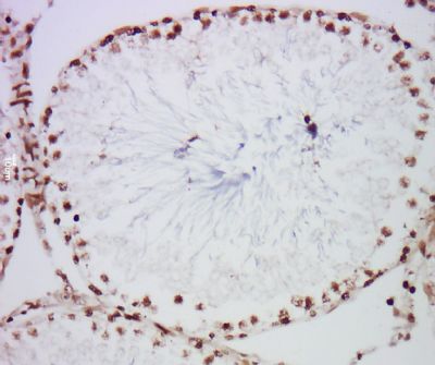 Tissue/cell: Rat testis tissue; 4% Paraformaldehyde-fixed and paraffin-embedded; Antigen retrieval: citrate buffer ( 0.01M, pH 6.0 ), Boiling bathing for 15min; Block endogenous peroxidase by 3% Hydrogen peroxide for 30min; Blocking buffer (normal goat serum,C-0005) at 37℃ for 20 min; Incubation: Anti-PEG3 Polyclonal Antibody, Unconjugated(bs-1870R) 1:200, overnight at 4°C, followed by conjugation to the secondary antibody(SP-0023) and DAB(C-0010) staining Tissue/cell: rat brain tissue; 4% Paraformaldehyde-fixed and paraffin-embedded; Antigen retrieval: citrate buffer ( 0.01M, pH 6.0 ), Boiling bathing for 15min; Block endogenous peroxidase by 3% Hydrogen peroxide for 30min; Blocking buffer (normal goat serum,C-0005) at 37℃ for 20 min; Incubation: Anti-PEG3 Polyclonal Antibody, Unconjugated(bs-1870R) 1:200, overnight at 4°C, followed by conjugation to the secondary antibody(SP-0023) and DAB(C-0010) staining  Blank control(black line):SH-SY5Y. Primary Antibody (green line): Rabbit Anti-PEG3 antibody (bs-1870R) Dilution:1ug/Test; Secondary Antibody : Goat anti-rabbit IgG-AF488 Dilution: 0.5ug/Test. Negative control(white blue line): PBS Isotype control(orange line): Normal Rabbit IgG Protocol The cells were fixed with 4% PFA (10min at room temperature)and then permeabilized with 90% ice-cold methanol for 20 min at -20℃.The cells were then incubated in 5%BSA to block non-specific protein-protein interactions for 30 min at room temperature .Cells stained with Primary Antibody for 30 min at room temperature. The secondary antibody used for 40 min at room temperature. Acquisition of 20,000 events was performed. |


