
上海细胞库
人源细胞系| 稳转细胞系| 基因敲除株| 基因点突变细胞株| 基因过表达细胞株| 重组细胞系| 猪的细胞系| 马细胞系| 兔的细胞系| 犬的细胞系| 山羊的细胞系| 鱼的细胞系| 猴的细胞系| 仓鼠的细胞系| 狗的细胞系| 牛的细胞| 大鼠细胞系| 小鼠细胞系| 其他细胞系|
人源细胞系| 稳转细胞系| 基因敲除株| 基因点突变细胞株| 基因过表达细胞株| 重组细胞系| 猪的细胞系| 马细胞系| 兔的细胞系| 犬的细胞系| 山羊的细胞系| 鱼的细胞系| 猴的细胞系| 仓鼠的细胞系| 狗的细胞系| 牛的细胞| 大鼠细胞系| 小鼠细胞系| 其他细胞系|

| 规格 | 价格 | 库存 |
|---|---|---|
| 50UL | ¥ 1200 | 6 |
| 100UL | ¥ 1900 | 5 |
| 200UL | ¥ 2900 | 2 |
| 产品编号 | Ys-7362R |
| 英文名称 | Melan A |
| 中文名称 | 黑色素瘤相关抗原/黑色素-A抗体 |
| 别 名 | MAR1_HUMAN; Melanoma antigen recognized by T-cells 1; MLANA; MART1; Antigen LB39-AA; Antigen SK29-AA; Protein Melan-A; |
| 研究领域 | 肿瘤 细胞生物 免疫学 t-淋巴细胞 |
| 抗体来源 | Rabbit |
| 克隆类型 | Polyclonal |
| 交叉反应 | Human, Mouse, (predicted: Rat, ) |
| 产品应用 | WB=1:500-2000 ELISA=1:5000-10000 IHC-P=1:100-500 IHC-F=1:100-500 ICC=1:100-500 IF=1:100-500 (石蜡切片需做抗原修复) not yet tested in other applications. optimal dilutions/concentrations should be determined by the end user. |
| 理论分子量 | 13kDa |
| 细胞定位 | 细胞浆 |
| 性 状 | Liquid |
| 浓 度 | 1mg/ml |
| 免 疫 原 | KLH conjugated synthetic peptide derived from mouse Melan A: 1-80/113 |
| 亚 型 | IgG |
| 纯化方法 | affinity purified by Protein A |
| 缓 冲 液 | 0.01M TBS(pH7.4) with 1% BSA, 0.03% Proclin300 and 50% Glycerol. |
| 保存条件 | Shipped at 4℃. Store at -20 °C for one year. Avoid repeated freeze/thaw cycles. |
| 注意事项 | This product as supplied is intended for research use only, not for use in human, therapeutic or diagnostic applications. |
| PubMed | PubMed |
| 产品介绍 | Melanoma-associated antigens recognized by cytotoxic T lymphocytes (CTL) have been grouped into three categories: melanocyte differentiation antigens, cancer/testis-specific antigens and mutated or aberrantly expressed antigens. Many of these antigens consist of peptides that are presented to T cells by HLA molecules; they represent potential targets for cancer immunotherapy. Melan-A (also designated MART-1) is a melanocyte differentiation antigen that is specific to melanomas, melanocyte cell lines and retina. Melan-A peptide is recognized by most HLA-A2-restricted tumor-specific tumor-infiltrating lymphocytes in patients with melanoma. Antimelanoma cytotoxic T lymphocytes can be generated with a Melan-A peptide, implicating Melan-A as a potential candidate for antigen-specific immunotherapy in melanoma patients. Function: Involved in melanosome biogenesis by ensuring the stability of GPR143. Plays a vital role in the expression, stability, trafficking, and processing of melanocyte protein PMEL, which is critical to the formation of stage II melanosomes. Subunit: Interacts with PMEL. Interacts with GPR143. Subcellular Location: Endoplasmic reticulum membrane. Golgi apparatus. Golgi apparatus > trans-Golgi network membrane. Melanosome. Also found in small vesicles and tubules dispersed over the entire cytoplasm. A small fraction of the protein is inserted into the membrane in an inverted orientation. Inversion of membrane topology results in the relocalization of the protein from a predominant Endoplasmic reticulum membrane. Golgi apparatus. Golgi apparatus > trans-Golgi network membrane. Melanosome. Also found in small vesicles and tubules dispersed over the entire cytoplasm. A small fraction of the protein is inserted into the membrane in an inverted orientation. Inversion of membrane topology results in the relocalization of the protein from a predominant Golgi/post-Golgi area to the endoplasmic reticulum. Melanoma cells expressing the protein with an inverted membrane topology are more effectively recognized by specific cytolytic T-lymphocytes than those expressing the protein in its native membrane orientation. Tissue Specificity: Expression is restricted to melanoma and melanocyte cell lines and retina. Post-translational modifications: Acylated. SWISS: Q2TA50 Gene ID: 77836 t |
| 产品图片 | 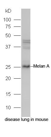 Protein: disease lung in mouse lysate; Primary: rabbit Anti-Melan A (bs-7362R) at 1:300; Secondary: HRP conjugated Goat-Anti-rabbit IgG(bs-0295G-HRP) at 1: 5000; Predicted band size: 26 kD Observed band size: 26 kD 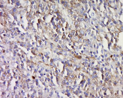 Tissue/cell: human melanoma tissue; 4% Paraformaldehyde-fixed and paraffin-embedded; Antigen retrieval: citrate buffer ( 0.01M, pH 6.0 ), Boiling bathing for 15min; Block endogenous peroxidase by 3% Hydrogen peroxide for 30min; Blocking buffer (normal goat serum,C-0005) at 37℃ for 20 min; Incubation: Anti-Melan A Polyclonal Antibody, Unconjugated(bs-7362R) 1:500, overnight at 4°C, followed by conjugation to the secondary antibody(SP-0023) and DAB(C-0010) staining |


