
上海细胞库
人源细胞系| 稳转细胞系| 基因敲除株| 基因点突变细胞株| 基因过表达细胞株| 重组细胞系| 猪的细胞系| 马细胞系| 兔的细胞系| 犬的细胞系| 山羊的细胞系| 鱼的细胞系| 猴的细胞系| 仓鼠的细胞系| 狗的细胞系| 牛的细胞| 大鼠细胞系| 小鼠细胞系| 其他细胞系|
人源细胞系| 稳转细胞系| 基因敲除株| 基因点突变细胞株| 基因过表达细胞株| 重组细胞系| 猪的细胞系| 马细胞系| 兔的细胞系| 犬的细胞系| 山羊的细胞系| 鱼的细胞系| 猴的细胞系| 仓鼠的细胞系| 狗的细胞系| 牛的细胞| 大鼠细胞系| 小鼠细胞系| 其他细胞系|

| 规格 | 价格 | 库存 |
|---|---|---|
| 50ul | ¥ 1100 | 6 |
| 100il | ¥ 1900 | 5 |
| 200ul | ¥ 2900 | 3 |
| 产品编号 | 8394R |
| 英文名称 | Fbxw7 |
| 中文名称 | Fbxw7蛋白抗体 |
| 别 名 | AGO; Archipelago homolog; Archipelago, Drosophila, homolog of antibody CDC4; DKFZp686F23254; F box and WD 40 domain protein 7 (archipelago homolog, Drosophila); F box and WD 40 domain protein 7; F box and WD repeat domain containing 7; F box protein FBW7; F box protein FBX30; F box protein SEL10; F-box and WD-40 domain-containing protein 7; F-box protein FBX30; F-box/WD repeat-containing protein 7; FBW6; FBW7; FBX30; FBXO30; FBXW6; FBXW7; FBXW7_HUMAN; FLJ16457; hAgo; hCdc4; Homolog of C elegans sel 10; Homolog of C.elegans sel10; SEL-10; SEL10. |
| 研究领域 | 肿瘤 细胞生物 免疫学 神经生物学 |
| 抗体来源 | Rabbit |
| 克隆类型 | Polyclonal |
| 交叉反应 | Human, Mouse, Rat, (predicted: Dog, Pig, Cow, Horse, ) |
| 产品应用 | ELISA=1:5000-10000 IHC-P=1:100-500 IHC-F=1:100-500 Flow-Cyt=2ug/Test ICC=1:100 IF=1:50-200 (石蜡切片需做抗原修复) not yet tested in other applications. optimal dilutions/concentrations should be determined by the end user. |
| 理论分子量 | 78kDa |
| 细胞定位 | 细胞核 细胞浆 |
| 性 状 | Liquid |
| 浓 度 | 1mg/ml |
| 免 疫 原 | KLH conjugated synthetic peptide derived from human Fbxw7/CDC4: 501-600/707 |
| 亚 型 | IgG |
| 纯化方法 | affinity purified by Protein A |
| 缓 冲 液 | 0.01M TBS(pH7.4) with 1% BSA, 0.03% Proclin300 and 50% Glycerol. |
| 保存条件 | Shipped at 4℃. Store at -20 °C for one year. Avoid repeated freeze/thaw cycles. |
| 注意事项 | This product as supplied is intended for research use only, not for use in human, therapeutic or diagnostic applications. |
| PubMed | PubMed |
| 产品介绍 | The F-box protein family is characterized by an approximately 40 amino acid motif known as the F-box. F-box proteins constitute one of the four subunits of ubiquitin protein ligase complex called SCFs (SKP1-cullin-F-box), which function in phosphorylation-dependent ubiquitination. One family member, Cdc4, also known as AGO, FBW7, FBXW7, FBX30, SEL10, and FLJ11071, maps to human chromosome 4q31.3. Alternative splicing of this gene generates four transcript variants. In addition to an F-box, Cdc4 contains seven tandem WD40 repeats. Cdc4 binds directly to cyclin E and targets cyclin E for ubiquitin-mediated degradation. Mutations of the Cdc4 gene are detected in ovarian and breast cancer cell lines, suggesting that the gene may be involved in the pathogenesis of human cancers. Function: Substrate recognition component of a SCF (SKP1-CUL1-F-box protein) E3 ubiquitin-protein ligase complex which mediates the ubiquitination and subsequent proteasomal degradation of target proteins. Probably recognizes and binds to phosphorylated target proteins. Involved in the degradation of cyclin-E, MYC, NOTCH1 released notch intracellular domain (NICD), and probably PSEN1. Subunit: Component of the SCF(FBXW7) complex consisting of CUL1, RBX1, SKP1 and FBXW7. Interacts with PSEN1, cyclin E, NOTCH1 NICD, NOTCH4 NICD and SKP1. Interacts with MYC (when phosphorylated). Isoform 1 interacts with USP28, leading to counteract ubiquitination of MYC. Isoform 4 interacts (via WD repeats) with SV40 large T antigen (via CPD region). Forms a trimeric complex with NOTCH1 and SGK1. Subcellular Location: Isoform 1: Nucleus, nucleoplasm. Isoform 2: Cytoplasm. Isoform 4: Nucleus, nucleolus. Nucleus. Tissue Specificity: Isoform 1 is widely expressed. Isoform 4 is expressed in brain. Post-translational modifications: Phosphorylated upon DNA damage, probably by ATM or ATR. Similarity: Contains 1 F-box domain. Contains 7 WD repeats. SWISS: Q969H0 Gene ID: 55294 |
| 产品图片 | 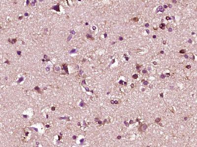 Paraformaldehyde-fixed, paraffin embedded (human brain glioma); Antigen retrieval by boiling in sodium citrate buffer (pH6.0) for 15min; Block endogenous peroxidase by 3% hydrogen peroxide for 20 minutes; Blocking buffer (normal goat serum) at 37°C for 30min; Antibody incubation with (Fbxw7) Polyclonal Antibody, Unconjugated (bs-8394R) at 1:400 overnight at 4°C, followed by operating according to SP Kit(Rabbit) (sp-0023) instructionsand DAB staining. 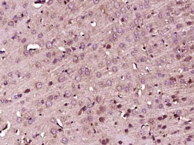 Paraformaldehyde-fixed, paraffin embedded (Mouse brain); Antigen retrieval by boiling in sodium citrate buffer (pH6.0) for 15min; Block endogenous peroxidase by 3% hydrogen peroxide for 20 minutes; Blocking buffer (normal goat serum) at 37°C for 30min; Antibody incubation with (Fbxw7) Polyclonal Antibody, Unconjugated (bs-8394R) at 1:400 overnight at 4°C, followed by operating according to SP Kit(Rabbit) (sp-0023) instructionsand DAB staining.  Tissue/cell: rat brain tissue; 4% Paraformaldehyde-fixed and paraffin-embedded; Antigen retrieval: citrate buffer ( 0.01M, pH 6.0 ), Boiling bathing for 15min; Block endogenous peroxidase by 3% Hydrogen peroxide for 30min; Blocking buffer (normal goat serum,C-0005) at 37℃ for 20 min; Incubation: Anti-Fbxw7/CDC4 Polyclonal Antibody, Unconjugated(bs-8394R) 1:200, overnight at 4°C, followed by conjugation to the secondary antibody(SP-0023) and DAB(C-0010) staining 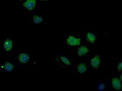 HepG2 cell; 4% Paraformaldehyde-fixed; Triton X-100 at room temperature for 20 min; Blocking buffer (normal goat serum, C-0005) at 37°C for 20 min; Antibody incubation with (Fbxw7) polyclonal Antibody, Unconjugated (bs-8394R) 1:100, 90 minutes at 37°C; followed by a conjugated Goat Anti-Rabbit IgG antibody at 37°C for 90 minutes, DAPI (blue, C02-04002) was used to stain the cell nuclei. 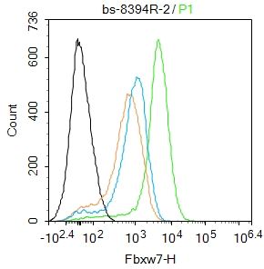 Blank control:Mouse spleen. Primary Antibody (green line): Rabbit Anti-Fbxw7 antibody (bs-8394R) Dilution: 2μg /10^6 cells; Isotype Control Antibody (orange line): Rabbit IgG . Secondary Antibody : Goat anti-rabbit IgG-FITC Dilution: 1μg /test. Protocol The cells were fixed with 4% PFA (10min at room temperature)and then permeabilized with 90% ice-cold methanol for 20 min at-20℃. The cells were then incubated in 5%BSA to block non-specific protein-protein interactions for 30 min at room temperature .Cells stained with Primary Antibody for 30 min at room temperature. The secondary antibody used for 40 min at room temperature. Acquisition of 20,000 events was performed. |


