
�Ϻ�ϸ����
��Դϸ��ϵ| ��תϸ��ϵ| �����ó���| �����ͻ��ϸ����| ���������ϸ����| ����ϸ��ϵ| ����ϸ��ϵ| ��ϸ��ϵ| �õ�ϸ��ϵ| Ȯ��ϸ��ϵ| ɽ���ϸ��ϵ| ���ϸ��ϵ| ���ϸ��ϵ| �����ϸ��ϵ| ����ϸ��ϵ| ţ��ϸ��| ����ϸ��ϵ| С��ϸ��ϵ| ����ϸ��ϵ|
��Դϸ��ϵ| ��תϸ��ϵ| �����ó���| �����ͻ��ϸ����| ���������ϸ����| ����ϸ��ϵ| ����ϸ��ϵ| ��ϸ��ϵ| �õ�ϸ��ϵ| Ȯ��ϸ��ϵ| ɽ���ϸ��ϵ| ���ϸ��ϵ| ���ϸ��ϵ| �����ϸ��ϵ| ����ϸ��ϵ| ţ��ϸ��| ����ϸ��ϵ| С��ϸ��ϵ| ����ϸ��ϵ|

| ��� | �۸� | ��� |
|---|---|---|
| 50ul | �� 1200 | 3 |
| 100ul | �� 1800.00 | 2 |
| �о����� | ϸ������ ����ѧ �ź�ת�� ��ϸ�� ϸ��ճ������ |
| ������Դ | Rabbit |
| ��¡���� | Polyclonal |
| ���淴Ӧ | Human, (predicted: Mouse, Pig, Cow, Rabbit, ) |
| ��ƷӦ�� | WB=1:500-2000 ELISA=1:5000-10000 IHC-P=1:100-500 IHC-F=1:100-500 Flow-Cyt=1μg/Test IF=1:100-500 ��ʯ����Ƭ������ԭ���� not yet tested in other applications. optimal dilutions/concentrations should be determined by the end user. |
| �� �� �� | 84kDa |
| ϸ����λ | ϸ��Ĥ |
| �� ״ | Liquid |
| Ũ �� | 1mg/ml |
| �� �� ԭ | KLH conjugated synthetic peptide derived from human Integrin beta 3:27-120/788 |
| �� �� | IgG |
| �������� | affinity purified by Protein A |
| �� �� Һ | 0.01M TBS(pH7.4) with 1% BSA, 0.03% Proclin300 and 50% Glycerol. |
| �������� | Shipped at 4��. Store at -20 °C for one year. Avoid repeated freeze/thaw cycles. |
| PubMed | PubMed |
| ��Ʒ���� | The ITGB3 (Integrin beta chain beta 3) protein product is the integrin beta chain beta 3. Integrins are integral cell-surface proteins composed of an alpha chain and a beta chain. A given chain may combine with multiple partners resulting in different integrins. Integrin beta 3 is found along with the alpha IIb chain in platelets. Integrins are known to participate in cell adhesion as well as cell-surface mediated signalling. Function: Integrin alpha-V/beta-3 is a receptor for cytotactin, fibronectin, laminin, matrix metalloproteinase-2, osteopontin, osteomodulin, prothrombin, thrombospondin, vitronectin and von Willebrand factor. Integrin alpha-IIb/beta-3 is a receptor for fibronectin, fibrinogen, plasminogen, prothrombin, thrombospondin and vitronectin. Integrins alpha-IIb/beta-3 and alpha-V/beta-3 recognize the sequence R-G-D in a wide array of ligands. Integrin alpha-IIb/beta-3 recognizes the sequence H-H-L-G-G-G-A-K-Q-A-G-D-V in fibrinogen gamma chain. Following activation integrin alpha-IIb/beta-3 brings about platelet/platelet interaction through binding of soluble fibrinogen. This step leads to rapid platelet aggregation which physically plugs ruptured endothelial surface. In case of HIV-1 infection, the interaction with extracellular viral Tat protein seems to enhance angiogenesis in Kaposi's sarcoma lesions. Subunit: Heterodimer of an alpha and a beta subunit. Beta-3 associates with either alpha-IIb or alpha-V. Isoform Beta-3C interacts with FLNB. Interacts with COMP. Interacts with HIV-1 Tat. Interacts with PDIA6 following platelet stimulation. Interacts with SYK; upon activation by ITGB3 promotes platelet adhesion. Interacts with MYO10. Subcellular Location: Membrane; Single-pass type I membrane protein. Tissue Specificity: Isoform beta-3A and isoform beta-3C are widely expressed. Isoform beta-3A is specifically expressed in osteoblast cells; isoform beta-3C is specifically expressed in prostate and testis. Post-translational modifications: Phosphorylated on tyrosine residues in response to thrombin-induced platelet aggregation. Probably involved in outside-in signaling. A peptide (AA 740-762) is capable of binding GRB2 only when both Tyr-773 and Tyr-785 are phosphorylated. Phosphorylation of Thr-779 inhibits SHC binding. DISEASE: Defects in ITGB3 are a cause of Glanzmann thrombasthenia (GT) [MIM:273800]; also known as thrombasthenia of Glanzmann and Naegeli. GT is the most common inherited disease of platelets. It is an autosomal recessive disorder characterized by mucocutaneous bleeding of mild-to-moderate severity and the inability of this integrin to recognize macromolecular or synthetic peptide ligands. GT has been classified clinically into types I and II. In type I, platelets show absence of the glycoprotein IIb/beta-3 complexes at their surface and lack fibrinogen and clot retraction capability. In type II, the platelets express the glycoprotein IIb/beta-3 complex at reduced levels (5-20% controls), have detectable amounts of fibrinogen, and have low or moderate clot retraction capability. The platelets of GT 'variants' have normal or near normal (60-100%) expression of dysfunctional receptors. Similarity: Belongs to the integrin beta chain family. Contains 1 VWFA domain. SWISS: P05106 Gene ID: 3690 Database links: Entrez Gene: 3690 Human Entrez Gene: 16416 Mouse Entrez Gene: 29302 Rat Omim: 173470 Human SwissProt: P05106 Human SwissProt: O54890 Mouse Unigene: 218040 Human Unigene: 87150 Mouse Important Note: This product as supplied is intended for research use only, not for use in human, therapeutic or diagnostic applications. CD61��ԭ�ֳ�ΪGP III a����һ�ֱ�����ѪС�塢��ϸ��������ϸ��������ϸ������Ƥϸ���ϵ��ǵ��ס�CD61��CD41����ѪС���ǵ���II b/III b�� |
| ��ƷͼƬ | 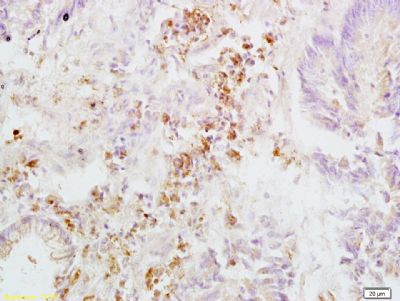 Tissue/cell: human rectal carcinoma; 4% Paraformaldehyde-fixed and paraffin-embedded; Antigen retrieval: citrate buffer ( 0.01M, pH 6.0 ), Boiling bathing for 15min; Block endogenous peroxidase by 3% Hydrogen peroxide for 30min; Blocking buffer (normal goat serum,C-0005) at 37�� for 20 min; Incubation: Anti-Integrin beta 3/CD61 Polyclonal Antibody, Unconjugated(Ys-0342R) 1:200, overnight at 4�C, followed by conjugation to the secondary antibody(SP-0023) and DAB(C-0010) staining 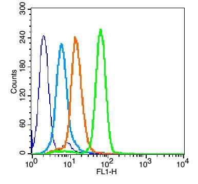 Overlay histogram showing HL 60 cells stained with Ys-0342R (Green line). The cells were fixed with 90% methanol (5 min) and then permeabilized with 0.01M PBS-Tween for 20 min. The cells were then incubated in 1x PBS / 10% normal goat serum to block non-specific protein-protein interactions followed by the antibody (bs-0342R,1μg/1x10^6 cells) for 30 min at 22��. The secondary antibody used was fluorescein isothiocyanate goat anti-rabbit IgG (H+L) (bs- 0295G-FITC , Brillant blue line) at 1/200 dilution for 30 min at 22��. Isotype control antibody was rabbit IgG (polyclonal,bs-0295P,Orange line) (1μg/1x10^6 cells) used under the same conditions. Unlabelled sample (blue line) was also used as a control. Acquisition of 20,000 events were collected using a 20mW Argon ion laser (488nm) and 525/30 bandpass filter. 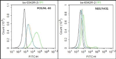 Black line : Positive blank control (HL60); Negative blank control (A431�� Green line : Primary Antibody (Rabbit Anti-CD61 antibody (Ys-0342R) ) Orange line��Isotype Control Antibody (Rabbit IgG) . Blue line : Secondary Antibody (Goat anti-rabbit IgG-AF488) HL60��Positive��and A431 Negative control��cells (black) were incubated in 5% BSA blocking buffer for 30 min at room temperature. Cells were then stained with CD61 Antibody(bs-0342R)at 1:50 dilution in blocking buffer and incubated for 30 min at room temperature, washed twice with 2% BSA in PBS, followed by secondary antibody(blue) incubation for 40 min at room temperature. Acquisitions of 20,000 events were performed. Cells stained with primary antibody (green), and isotype control (orange). |


