
上海细胞库
人源细胞系| 稳转细胞系| 基因敲除株| 基因点突变细胞株| 基因过表达细胞株| 重组细胞系| 猪的细胞系| 马细胞系| 兔的细胞系| 犬的细胞系| 山羊的细胞系| 鱼的细胞系| 猴的细胞系| 仓鼠的细胞系| 狗的细胞系| 牛的细胞| 大鼠细胞系| 小鼠细胞系| 其他细胞系|
人源细胞系| 稳转细胞系| 基因敲除株| 基因点突变细胞株| 基因过表达细胞株| 重组细胞系| 猪的细胞系| 马细胞系| 兔的细胞系| 犬的细胞系| 山羊的细胞系| 鱼的细胞系| 猴的细胞系| 仓鼠的细胞系| 狗的细胞系| 牛的细胞| 大鼠细胞系| 小鼠细胞系| 其他细胞系|

| 规格 | 价格 | 库存 |
|---|---|---|
| 50ul | ¥ 1600 | 200 |
| 100ul | ¥ 2600 | 200 |
中文名称 丝裂原活化蛋白激酶1/ERK 1/2重组兔单克隆抗体
| 产品图片 | 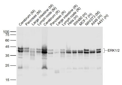 Sample: Lane 1: Cerebrum (Mouse) Lysate at 40 ug Lane 2: Pancreas (Mouse) Lysate at 40 ug Lane 3: Large intestine (Mouse) Lysate at 40 ug Lane 4: Lymph node (Mouse) Lysate at 40 ug Lane 5: Cerebrum (Rat) Lysate at 40 ug Lane 6: Pancreas (Rat) Lysate at 40 ug Lane 7: Large intestine (Rat) Lysate at 40 ug Lane 8: Lymph node (Rat) Lysate at 40 ug Lane 9: Hela (Human) Cell Lysate at 30 ug Lane 10: SW480 (Human) Cell Lysate at 30 ug Lane 11: MCF-7 (Human) Cell Lysate at 30 ug Lane 12: NIH/3T3 (Mouse) Cell Lysate at 30 ug Lane 13: A549 (Human) Cell Lysate at 30 ug Lane 14: A431 (Human) Cell Lysate at 30 ug Primary: Anti- ERK1/2 (bsm-52259R) at 1/1000 dilution Secondary: IRDye800CW Goat Anti-Rabbit IgG at 1/20000 dilution Predicted band size: 44/42 kD Observed band size: 44/42 kD  Paraformaldehyde-fixed, paraffin embedded (mouse brain); Antigen retrieval by boiling in sodium citrate buffer (pH6.0) for 15min; Block endogenous peroxidase by 3% hydrogen peroxide for 20 minutes; Blocking buffer (normal goat serum) at 37°C for 30min; Antibody incubation with (ERK1 2) Monoclonal Antibody, Unconjugated (bsm-52259R) at 1:200 overnight at 4°C, followed by operating according to SP Kit(Rabbit) (sp-0023) instructionsand DAB staining.  Paraformaldehyde-fixed, paraffin embedded (rat brain); Antigen retrieval by boiling in sodium citrate buffer (pH6.0) for 15min; Block endogenous peroxidase by 3% hydrogen peroxide for 20 minutes; Blocking buffer (normal goat serum) at 37°C for 30min; Antibody incubation with (ERK1 2) Monoclonal Antibody, Unconjugated (bsm-52259R) at 1:200 overnight at 4°C, followed by operating according to SP Kit(Rabbit) (sp-0023) instructionsand DAB staining. 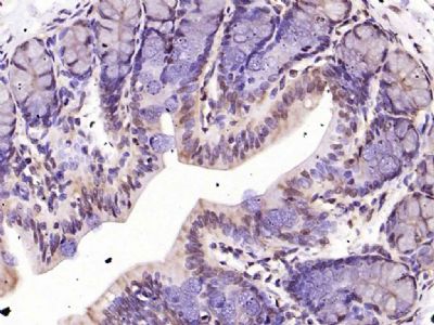 Paraformaldehyde-fixed, paraffin embedded (rat colon); Antigen retrieval by boiling in sodium citrate buffer (pH6.0) for 15min; Block endogenous peroxidase by 3% hydrogen peroxide for 20 minutes; Blocking buffer (normal goat serum) at 37°C for 30min; Antibody incubation with (ERK1 2) Monoclonal Antibody, Unconjugated (bsm-52259R) at 1:200 overnight at 4°C, followed by operating according to SP Kit(Rabbit) (sp-0023) instructionsand DAB staining. 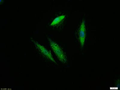 Tissue/cell:A549 cell;4% Paraformaldehyde-fixed;Triton X-100 at room temperature for 20 min; Blocking buffer (normal goat serum,C-0005) at 37°C for 20 min; Antibody incubation with (ERK1/2) monoclonal Antibody, Unconjugated (bsm-52259R) 1:100, 90 minutes at 37°C; followed by a FITC conjugated Goat Anti-Rabbit IgG antibody at 37°C for 90 minutes, DAPI (blue, C02-04002) was used to stain the cell nuclei.  Blank control: Hela. Primary Antibody (green line): Rabbit Anti-ERK1/2 antibody (bsm-52259R) Dilution: 1μg /10^6 cells; Isotype Control Antibody (orange line): Rabbit IgG . Secondary Antibody : Goat anti-rabbit IgG-AF647 Dilution: 1μg /test. Protocol The cells were fixed with 4% PFA (10min at room temperature)and then permeabilized with 90% ice-cold methanol for 20 min at -20℃. The cells were then incubated in 5%BSA to block non-specific protein-protein interactions for 30 min at room temperature .Cells stained with Primary Antibody for 30 min at room temperature. The secondary antibody used for 40 min at room temperature. Acquisition of 20,000 events was performed. |


