
上海细胞库
人源细胞系| 稳转细胞系| 基因敲除株| 基因点突变细胞株| 基因过表达细胞株| 重组细胞系| 猪的细胞系| 马细胞系| 兔的细胞系| 犬的细胞系| 山羊的细胞系| 鱼的细胞系| 猴的细胞系| 仓鼠的细胞系| 狗的细胞系| 牛的细胞| 大鼠细胞系| 小鼠细胞系| 其他细胞系|
人源细胞系| 稳转细胞系| 基因敲除株| 基因点突变细胞株| 基因过表达细胞株| 重组细胞系| 猪的细胞系| 马细胞系| 兔的细胞系| 犬的细胞系| 山羊的细胞系| 鱼的细胞系| 猴的细胞系| 仓鼠的细胞系| 狗的细胞系| 牛的细胞| 大鼠细胞系| 小鼠细胞系| 其他细胞系|

| 规格 | 价格 | 库存 |
|---|---|---|
| 50μl | ¥ 980 | 200 |
| 来源 | 用途 | 交叉反应性 | 理论分子量 | 实际分子量 |
| Rabbit | WB, IF, IHC | H, M, R | 66kDa | 70kDa |
WB, Western blot; IP, Immunoprecipitation; IF, Immunofluorescence; IHC, Immunohistochemistry; ICC, Immunocytochemistry; FC, Flow Cytometry; ChIP, Chromatin Immunoprecipitation Assay; ChIP-seq, ChIP-sequencing.
H, Human; M, Mouse; R, Rat; C, Chicken; Cw, Cow; Dg, Dog; Gp, Guinea pig; Hm, Hamster; Hr, Horse; Mk, Monkey; Pg, Pig; Rb, Rabbit; S, Sheep; Z, Zebrafish; All, all species expected.
配套提供了Western一抗稀释液,可以用于Western检测或其它适当用途时的一抗稀释。
建议抗体使用时的稀释比例如下(实际使用时需根据抗原水平的高低作适当调整):
| WB | IP | IF | IHC | ICC | FC | ChIP | ChIP-seq |
| 1:500-1:2000 | - | 1:50-1:200 | 1:50-1:200 | 1:50-1:200 | - | - | - |
抗体详细信息如下::
| About this Antibody | |
| Name | Lamin B1 Rabbit Polyclonal Antibody (KO Validated) |
| Category | Rabbit Polyclonal Antibody (pAb); Primary antibody |
| Isotype | IgG |
| Purification method | Affinity purification |
| Positive samples | HepG2, Jurkat, HT-29, U-251MG, A-549, Mouse spleen, Rat testis |
| Cellular location | Lipid-anchor, Nucleoplasmic side, Nucleus inner membrane |
| Customer validation | WB (Human, H M); IHC (Mouse); IP (H M) |
| About the Immunogen | |
| Immunogen | Recombinant fusion protein of human Lamin B1 (NP_005564.1). |
| Sequence | RVTVSRASSSRSVRTTRGKRKRVDVEESEASSSVSISHSASATGNVCIEEIDVDGKFIRLKNTSEQDQPMGGWEMIRKIGDTSVSYKYTSRYVLKAGQTVTIWAANAGVTASPPTDLIWKNQNSWGTGEDVKVILKNSQGEEVAQRSTVFKTTIPEEEEEEEEAAGVVVEEELFHQQGTPRASNRSCAIM |
| Gene ID | 4001 |
| Swiss Prot | P20700 |
| Synonyms | LMNB1; ADLD; LMN; LMN2; LMNB; lamin-B1 |
| Category | Death Receptor Signaling; PI3K/Akt Signaling |
| Background | Lamins are nuclear membrane structural components that are important in maintaining normal cell functions, such as cell cycle control, DNA replication, and chromatin organization. Lamins have been subdivided into types A and B. Type-A lamins consist of lamin A and C, which arise from alternative splicing of the lamin A gene LMNA. Lamin A and C are cleaved by caspases into large (41-50 kDa) and small (28 kDa) fragments, which can be used as markers for apoptosis. Type-B lamins consist of lamin B1 and B2, encoded by separate genes. Lamin B1 is also cleaved by caspases during apoptosis. Research studies have shown that duplication of the lamin B1 gene LMNB1 is correlated with pathogenesis of the neurological disorder adult-onset leukodystrophy. |
包装清单:
| 产品编号 | 产品名称 | 包装 |
| AF5222 | Lamin B1 Rabbit Polyclonal Antibody (KO Validated) | 50μl |
| AZ050 | Western一抗稀释液 | 50ml |
| — | 说明书 | 1份 |
保存条件:
Lamin B1 Rabbit Polyclonal Antibody (KO Validated) -20ºC保存,Western一抗稀释液-20ºC或4ºC保存,一年有效。Western一抗稀释液优先推荐4ºC保存,长期不使用可以考虑-20ºC保存,但冻融可能会导致出现轻微的浑浊和少量不溶物。
注意事项:
如果本抗体用于Western blot (WB)、免疫荧光(IF)、免疫细胞化学(ICC)等实验,请注意回收使用过的稀释抗体。回收的抗体通常至少可以重复使用5-10次。稀释后的抗体,包括已经使用过的稀释抗体,请4℃保存。
回收后重复使用的抗体,使用方法同新鲜稀释的抗体。如果在重复使用过程中发现抗体出现轻微混浊现象,可以10,000g离心1-3分钟,取上清用于后续检测。如果回收的抗体出现明显的絮状物或长霉长菌等情况,则可以考虑废弃该抗体。
提供的Western一抗稀释液也可以用于免疫荧光(IF)、免疫组化(IHC)、免疫细胞化学(ICC)等适当用途。如果希望获得最佳的检测效果,请考虑使用上述检测专用的一抗稀释液。
本产品仅限于专业人员的科学研究用,不得用于临床诊断或治疗,不得用于食品或药品,不得存放于普通住宅内。
为了您的安全和健康,请穿实验服并戴一次性手套操作。
3. 其它实验操作请自行参考适当的protocol进行。
4. 代表性图片:
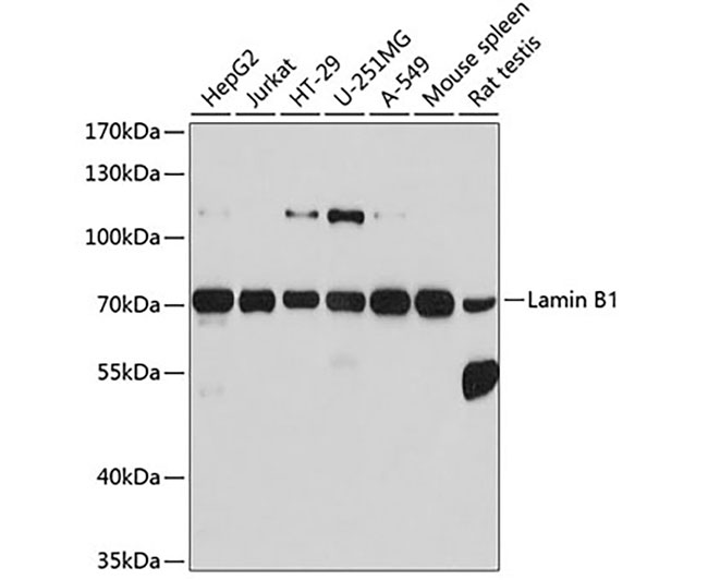 | 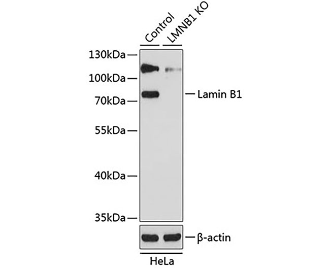 |
| Fig. 1. Western blot analysis of extracts of various cell lines, using Lamin B1 antibody at 1:1000 dilution. Secondary antibody: HRP-labeled Goat Anti-Rabbit IgG(H+L) (A0208) at 1:1000 dilution. Lysates/proteins: 25µg per lane. Blocking buffer: QuickBlock™ Blocking Buffer (P0231). Detection: BeyoECL Star (P0018A). Exposure time:1s. | Fig. 2. Western blot analysis of extracts from normal (control) and Lamin B1 knockout (KO) HeLa cells, using Lamin B1 antibody at 1:1000 dilution. Secondary antibody: HRP-labeled Goat Anti-Rabbit IgG(H+L) (A0208) at 1:1000 dilution. Lysates/proteins: 25µg per lane. Blocking buffer: QuickBlock™ Blocking Buffer (P0231). Detection: BeyoECL Star (P0018A). Exposure time:1s. |
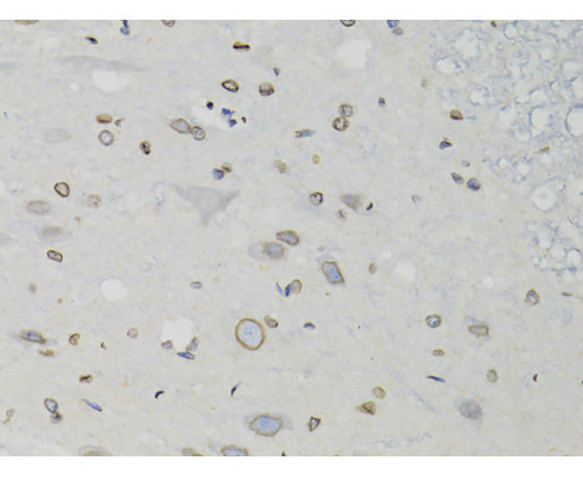 | 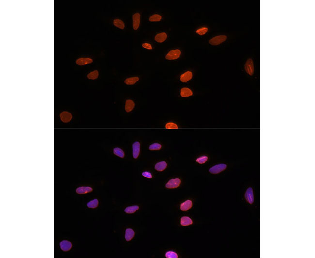 |
| Fig. 3. Immunohistochemistry of paraffin-embedded rat spinal cord using Lamin B1 antibody at dilution of 1:100 (40x lens). | Fig. 4. Immunofluorescence analysis of U-2 OS cells using Lamin B1 Polyclonal Antibody at dilution of 1:100 (40x lens). Blue: DAPI for nuclear staining. |


