
上海细胞库
人源细胞系| 稳转细胞系| 基因敲除株| 基因点突变细胞株| 基因过表达细胞株| 重组细胞系| 猪的细胞系| 马细胞系| 兔的细胞系| 犬的细胞系| 山羊的细胞系| 鱼的细胞系| 猴的细胞系| 仓鼠的细胞系| 狗的细胞系| 牛的细胞| 大鼠细胞系| 小鼠细胞系| 其他细胞系|
人源细胞系| 稳转细胞系| 基因敲除株| 基因点突变细胞株| 基因过表达细胞株| 重组细胞系| 猪的细胞系| 马细胞系| 兔的细胞系| 犬的细胞系| 山羊的细胞系| 鱼的细胞系| 猴的细胞系| 仓鼠的细胞系| 狗的细胞系| 牛的细胞| 大鼠细胞系| 小鼠细胞系| 其他细胞系|

| 规格 | 价格 | 库存 |
|---|---|---|
| 50μl | ¥ 980 | 200 |
| 来源 | 用途 | 交叉反应性 | 理论分子量 | 实际分子量 |
| Rabbit | WB, IF | H, M | 54/117kDa | 118kDa |
WB, Western blot; IP, Immunoprecipitation; IF, Immunofluorescence; IHC, Immunohistochemistry; ICC, Immunocytochemistry; FC, Flow Cytometry; ChIP, Chromatin Immunoprecipitation Assay; ChIP-seq, ChIP-sequencing.
H, Human; M, Mouse; R, Rat; C, Chicken; Cw, Cow; Dg, Dog; Gp, Guinea pig; Hm, Hamster; Hr, Horse; Mk, Monkey; Pg, Pig; Rb, Rabbit; S, Sheep; Z, Zebrafish; All, all species expected.
配套提供了Western一抗稀释液,可以用于Western检测或其它适当用途时的一抗稀释。
建议抗体使用时的稀释比例如下(实际使用时需根据抗原水平的高低作适当调整):
| WB | IP | IF | IHC | ICC | FC | ChIP | ChIP-seq |
| 1:500-1:2000 | - | 1:10-1:100 | - | - | - | - | - |
抗体详细信息如下::
| About this Antibody | |
| Name | IDE/Insulysin Rabbit Polyclonal Antibody (KO Validated) |
| Category | Rabbit Polyclonal Antibody (pAb); Primary antibody |
| Isotype | IgG |
| Purification method | Affinity purification |
| Positive samples | DU145, Mouse liver |
| Cellular location | Cell membrane, Cytoplasm, Secreted |
| Customer validation | - |
| About the Immunogen | |
| Immunogen | Recombinant fusion protein of human IDE (NP_004960.2). |
| Sequence | MRYRLAWLLHPALPSTFRSVLGARLPPPERLCGFQKKTYSKMNNPAIKRIGNHITKSPEDKREYRGLELANGIKVLLISDPTTDKSSAALDVHIGSLSDPPNIAGLSHFCEHMLFLGTKKYPKENEYSQFLSEHAGSSNAFTSGEHTNYYFDVSHEHLEGALDRFAQFFLCPLFDESCKDREVNAVDSEHEKNVMNDAWRLFQLEKATGNPKHPFSKFGTGNKYTLETRPNQEGIDVRQELLKFHSAYYS |
| Gene ID | 3416 |
| Swiss Prot | P14735 |
| Synonyms | IDE; INSULYSIN; insulin-degrading enzyme |
| Category | Insulin Signaling; Amyloid Plaque and Neurofibrillary Tangle Formation in Alzheimer's Disease |
| Background | Insulysin, or insulin-degrading enzyme (IDE), is a zinc metallopeptidase of the inverzincin family. IDE is primarily located in the cytosol, but has been detected as a secreted enzyme and associated with the plasma membrane as well. The enzyme is expressed in many tissues, with the highest levels in liver, kidney, brain, and testis. IDE hydrolyzes a variety of regulatory peptides, including insulin, glucagon, atrial natriuretic factor, and transforming growth factor-alpha in vitro. In addition, IDE has been shown to degrade the amyloid beta (A beta ) peptide, which polymerizes into the plaques associated with Alzheimer's disease. Deficiencies in IDE activity may contribute to the pathogenesis of type 2 diabetes mellitus (DM2) and Alzheimer's disease. The IDE region of human chromosome 10q has been genetically linked to DM2. When the IDE gene was specifically disrupted in mice, IDE -/- animals developed hyperinsulinemia and glucose intolerance, characteristics of DM2. The IDE -/- mice were also shown to have a significant decrease in A beta degradation in the brain, resulting in increased cerebral accumulation of A beta peptide. This in vivo evidence is consistent with the hypotheses that IDE is important for the degradation of insulin in cells and for the clearance of A beta peptide in the brain. |
包装清单:
| 产品编号 | 产品名称 | 包装 |
| AF5201 | IDE/Insulysin Rabbit Polyclonal Antibody (KO Validated) | 50μl |
| AZ050 | Western一抗稀释液 | 50ml |
| — | 说明书 | 1份 |
保存条件:
IDE/Insulysin Rabbit Polyclonal Antibody (KO Validated) -20ºC保存,Western一抗稀释液-20ºC或4ºC保存,一年有效。Western一抗稀释液优先推荐4ºC保存,长期不使用可以考虑-20ºC保存,但冻融可能会导致出现轻微的浑浊和少量不溶物。
注意事项:
如果本抗体用于Western blot (WB)、免疫荧光(IF)、免疫细胞化学(ICC)等实验,请注意回收使用过的稀释抗体。回收的抗体通常至少可以重复使用5-10次。稀释后的抗体,包括已经使用过的稀释抗体,请4℃保存。
回收后重复使用的抗体,使用方法同新鲜稀释的抗体。如果在重复使用过程中发现抗体出现轻微混浊现象,可以10,000g离心1-3分钟,取上清用于后续检测。如果回收的抗体出现明显的絮状物或长霉长菌等情况,则可以考虑废弃该抗体。
提供的Western一抗稀释液也可以用于免疫荧光(IF)、免疫组化(IHC)、免疫细胞化学(ICC)等适当用途。如果希望获得最佳的检测效果,请考虑使用上述检测专用的一抗稀释液。
本产品仅限于专业人员的科学研究用,不得用于临床诊断或治疗,不得用于食品或药品,不得存放于普通住宅内。
为了您的安全和健康,请穿实验服并戴一次性手套操作。
3. 其它实验操作请自行参考适当的protocol进行。
4. 代表性图片:
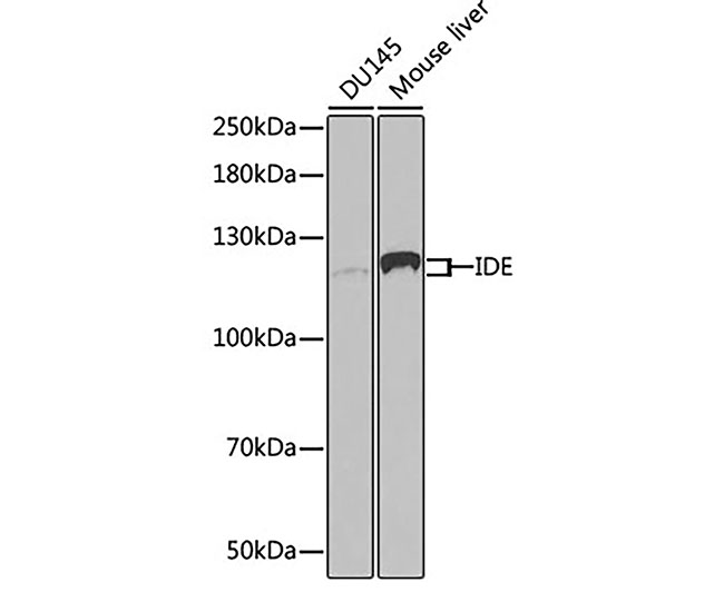 | 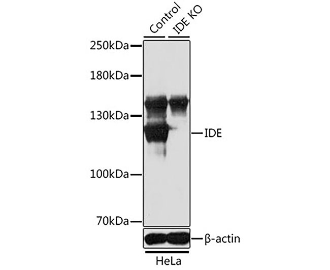 |
| Fig. 1. Western blot analysis of extracts of various cell lines, using IDE antibody at 1:1000 dilution. Secondary antibody: HRP-labeled Goat Anti-Rabbit IgG(H+L) (A0208) at 1:1000 dilution. Lysates/proteins: 25µg per lane. Blocking buffer: QuickBlock™ Blocking Buffer (P0231). Detection: BeyoECL Star (P0018A). | Fig. 2. Western blot analysis of extracts from normal (control) and IDE knockout (KO) HeLa cells, using IDE antibody at 1:1000 dilution. Secondary antibody: HRP-labeled Goat Anti-Rabbit IgG(H+L) (A0208) at 1:1000 dilution. Lysates/proteins: 25µg per lane. Blocking buffer: QuickBlock™ Blocking Buffer (P0231). Detection: BeyoECL Star (P0018A). Exposure time:1s. |
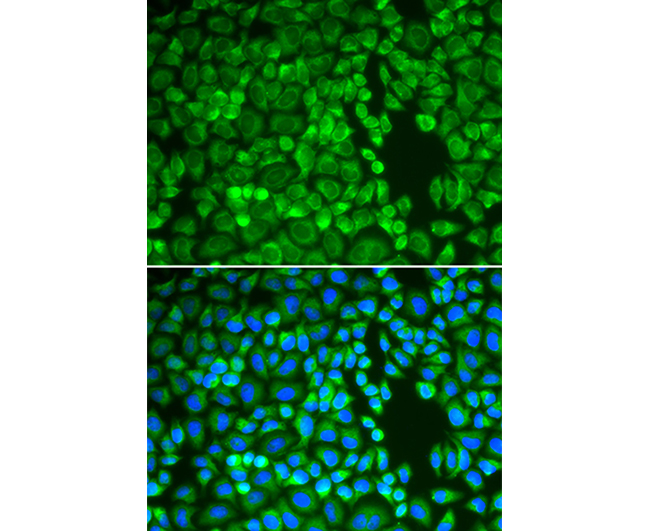 | |
| Fig. 3. Immunofluorescence analysis of A549 cells using IDE antibody. Blue: DAPI for nuclear staining. | |


