
上海细胞库
人源细胞系| 稳转细胞系| 基因敲除株| 基因点突变细胞株| 基因过表达细胞株| 重组细胞系| 猪的细胞系| 马细胞系| 兔的细胞系| 犬的细胞系| 山羊的细胞系| 鱼的细胞系| 猴的细胞系| 仓鼠的细胞系| 狗的细胞系| 牛的细胞| 大鼠细胞系| 小鼠细胞系| 其他细胞系|
人源细胞系| 稳转细胞系| 基因敲除株| 基因点突变细胞株| 基因过表达细胞株| 重组细胞系| 猪的细胞系| 马细胞系| 兔的细胞系| 犬的细胞系| 山羊的细胞系| 鱼的细胞系| 猴的细胞系| 仓鼠的细胞系| 狗的细胞系| 牛的细胞| 大鼠细胞系| 小鼠细胞系| 其他细胞系|

| 规格 | 价格 | 库存 |
|---|---|---|
| 50μl | ¥ 980 | 200 |
| 来源 | 用途 | 交叉反应性 | 理论分子量 | 实际分子量 |
| Rabbit | WB, IF | H, M, R | 3/15/32-53/73-81kDa | 82kDa |
WB, Western blot; IP, Immunoprecipitation; IF, Immunofluorescence; IHC, Immunohistochemistry; ICC, Immunocytochemistry; FC, Flow Cytometry; ChIP, Chromatin Immunoprecipitation Assay; ChIP-seq, ChIP-sequencing.
H, Human; M, Mouse; R, Rat; C, Chicken; Cw, Cow; Dg, Dog; Gp, Guinea pig; Hm, Hamster; Hr, Horse; Mk, Monkey; Pg, Pig; Rb, Rabbit; S, Sheep; Z, Zebrafish; All, all species expected.
配套提供了Western一抗稀释液,可以用于Western检测或其它适当用途时的一抗稀释。
建议抗体使用时的稀释比例如下(实际使用时需根据抗原水平的高低作适当调整):
| WB | IP | IF | IHC | ICC | FC | ChIP | ChIP-seq |
| 1:500-1:2000 | - | 1:50-1:200 | - | - | - | - | - |
抗体详细信息如下::
| About this Antibody | |
| Name | CD44 Rabbit Polyclonal Antibody (KO Validated) |
| Category | Rabbit Polyclonal Antibody (pAb); Primary antibody |
| Isotype | IgG |
| Purification method | Affinity purification |
| Positive samples | Mouse spleen |
| Cellular location | Cell membrane, Single-pass type I membrane protein |
| Customer validation | - |
| About the Immunogen | |
| Immunogen | Recombinant fusion protein of human CD44 (NP_001189486.1). |
| Sequence | VNRDGTRYVQKGEYRTNPEDIYPSNPTDDDVSSGSSSERSSTSGGYIFYTFSTVHPIPDEDSPWITDSTDRIPATRDQDTFHPSGGSHTTHGSESDGHSH |
| Gene ID | 960 |
| Swiss Prot | P16070 |
| Synonyms | CD44; CDW44; CSPG8; ECMR-III; HCELL; HUTCH-I; IN; LHR; MC56; MDU2; MDU3; MIC4; Pgp1; CD44 antigen |
| Category | Hippo Signaling Pathway |
| Background | CD44 is a type I transmembrane glycoprotein that mediates cell-cell and cell-matrix interaction through its affinity for hyaluronic acid (HA) and possibly through other parts of the extracellular matrix (ECM). CD44 is highly polymorphic, possesses a number of alternative splice variants and undergoes extensive post-translational modifications. Increased surface levels of CD44 are characteristic of T cell activation, and expression of the protein is upregulated during the inflammatory response. Research studies have shown that interactions between CD44 and HER2 are linked to an increase in ovarian carcinoma cell growth. CD44 interacts with ezrin, radixin and moesin (ERM), linking the actin cytoskeleton to the plasma membrane and the ECM. CD44 is constitutively phosphorylated at Ser325 in resting cells. Activation of PKC results in phosphorylation of Ser291, dephosphorylation of Ser325, disassociation of ezrin from CD44, and directional motility. |
包装清单:
| 产品编号 | 产品名称 | 包装 |
| AF5138 | CD44 Rabbit Polyclonal Antibody (KO Validated) | 50μl |
| AZ050 | Western一抗稀释液 | 50ml |
| — | 说明书 | 1份 |
保存条件:
CD44 Rabbit Polyclonal Antibody (KO Validated) -20ºC保存,Western一抗稀释液-20ºC或4ºC保存,一年有效。Western一抗稀释液优先推荐4ºC保存,长期不使用可以考虑-20ºC保存,但冻融可能会导致出现轻微的浑浊和少量不溶物。
注意事项:
如果本抗体用于Western blot (WB)、免疫荧光(IF)、免疫细胞化学(ICC)等实验,请注意回收使用过的稀释抗体。回收的抗体通常至少可以重复使用5-10次。稀释后的抗体,包括已经使用过的稀释抗体,请4℃保存。
回收后重复使用的抗体,使用方法同新鲜稀释的抗体。如果在重复使用过程中发现抗体出现轻微混浊现象,可以10,000g离心1-3分钟,取上清用于后续检测。如果回收的抗体出现明显的絮状物或长霉长菌等情况,则可以考虑废弃该抗体。
提供的Western一抗稀释液也可以用于免疫荧光(IF)、免疫组化(IHC)、免疫细胞化学(ICC)等适当用途。如果希望获得最佳的检测效果,请考虑使用上述检测专用的一抗稀释液。
本产品仅限于专业人员的科学研究用,不得用于临床诊断或治疗,不得用于食品或药品,不得存放于普通住宅内。
为了您的安全和健康,请穿实验服并戴一次性手套操作。
3. 其它实验操作请自行参考适当的protocol进行。
4. 代表性图片:
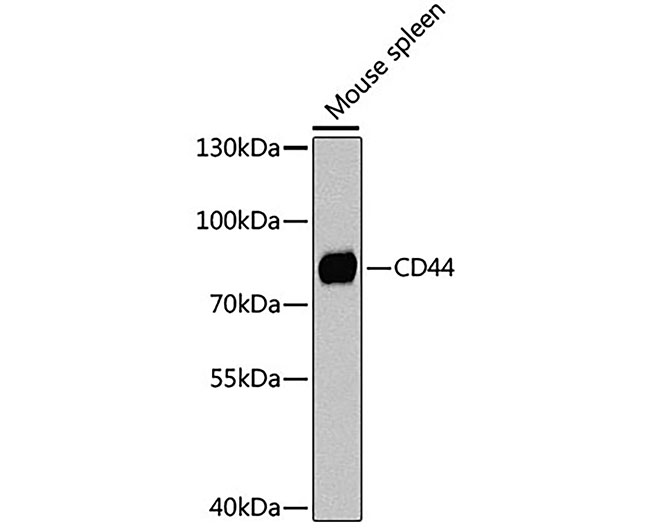 | 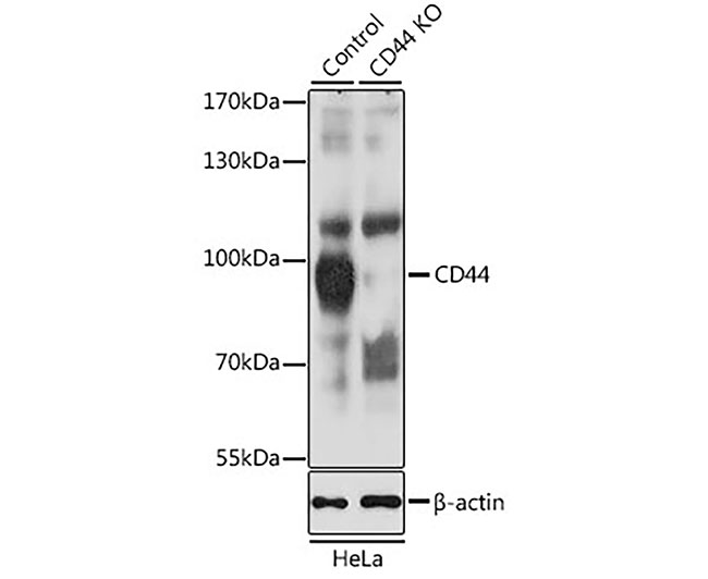 |
| Fig. 1. Western blot analysis of extracts of mouse spleen, using CD44 Antibody at 1:1000 dilution. Secondary antibody: HRP-labeled Goat Anti-Rabbit IgG(H+L) (A0208) at 1:1000 dilution. Lysates/proteins: 25µg per lane. Blocking buffer: QuickBlock™ Blocking Buffer (P0231). Detection: BeyoECL Star (P0018A). Exposure time:1s. | Fig. 2. Western blot analysis of extracts from normal (control) and CD44 knockout (KO) HeLa cells, using CD44 antibody at 1:1000 dilution. Secondary antibody: HRP-labeled Goat Anti-Rabbit IgG(H+L) (A0208) at 1:1000 dilution. Lysates/proteins: 25µg per lane. Blocking buffer: QuickBlock™ Blocking Buffer (P0231). Detection: BeyoECL Star (P0018A). Exposure time:1s. |
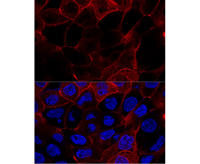 | 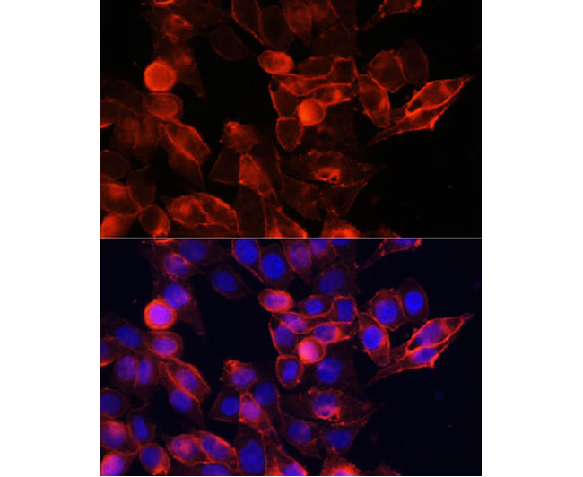 |
| Fig. 3. Immunofluorescence analysis of HeLa cells using CD44 antibody at dilution of 1:100. Blue: DAPI for nuclear staining. | Fig. 4. Confocal immunofluorescence analysis of A431 cells using CD44 antibody at dilution of 1:200. Blue: DAPI for nuclear staining. |


