
上海细胞库
人源细胞系| 稳转细胞系| 基因敲除株| 基因点突变细胞株| 基因过表达细胞株| 重组细胞系| 猪的细胞系| 马细胞系| 兔的细胞系| 犬的细胞系| 山羊的细胞系| 鱼的细胞系| 猴的细胞系| 仓鼠的细胞系| 狗的细胞系| 牛的细胞| 大鼠细胞系| 小鼠细胞系| 其他细胞系|
人源细胞系| 稳转细胞系| 基因敲除株| 基因点突变细胞株| 基因过表达细胞株| 重组细胞系| 猪的细胞系| 马细胞系| 兔的细胞系| 犬的细胞系| 山羊的细胞系| 鱼的细胞系| 猴的细胞系| 仓鼠的细胞系| 狗的细胞系| 牛的细胞| 大鼠细胞系| 小鼠细胞系| 其他细胞系|

| 规格 | 价格 | 库存 |
|---|---|---|
| 50ul | ¥ 980 | 200 |
| 100ul | ¥ 1680 | 200 |
| 200ul | ¥ 2480 | 200 |
| 中文名称 | 血小板内皮细胞黏附分子-1抗体 |
| 别 名 | platelet endothelial cell adhesion molecule precursor-1; PECAM-1; PECAM1; Adhesion molecule; CD31 antigen; CD31 EndoCAM; Endocam; FLJ34100; FLJ58394; GPIIA; Pecam 1; PECA1_HUMAN; PECAM 1 CD31 EndoCAM; PECA1; Pecam1; Platelet endothelial cell adhesion molecule; Platelet/endothelial cell adhesion molecule 1; Adhesion molecule; Platelet/endothelial cell adhesion molecule |
| 研究领域 | 肿瘤 心血管 细胞生物 免疫学 信号转导 干细胞 细胞粘附分子 细胞表面分子 糖蛋白 淋巴细胞 t-淋巴细胞 骨髓细胞 |
| 抗体来源 | Rabbit |
| 克隆类型 | Polyclonal |
| 交叉反应 | Human, Dog, Pig, (predicted: Mouse, Rat, Horse, ) |
| 产品应用 | WB=1:500-2000 ELISA=1:500-1000 Flow-Cyt=1μg/Test not yet tested in other applications. optimal dilutions/concentrations should be determined by the end user. |
| 分 子 量 | 78kDa |
| 细胞定位 | 细胞膜 |
| 性 状 | Liquid |
| 浓 度 | 1mg/ml |
| 免 疫 原 | KLH conjugated synthetic peptide derived from human CD31:601-680/738 |
| 亚 型 | IgG |
| 纯化方法 | affinity purified by Protein A |
| 储 存 液 | 0.01M TBS(pH7.4) with 1% BSA, 0.03% Proclin300 and 50% Glycerol. |
| 保存条件 | Shipped at 4℃. Store at -20 °C for one year. Avoid repeated freeze/thaw cycles. |
| PubMed | PubMed |
| 产品介绍 | Platelet endothelial cell adhesion molecule (PECAM-1) also known as cluster of differentiation 31 (CD31) is a protein that in humans is encoded by the PECAM1 gene found on chromosome 17. PECAM-1 plays a key role in removing aged neutrophils from the body. PECAM-1 is found on the surface of platelets, monocytes, neutrophils, and some types of T-cells, and makes up a large portion of endothelial cell intercellular junctions. The encoded protein is a member of the immunoglobulin superfamily and is likely involved in leukocyte transmigration, angiogenesis, and integrin activation. Function: Induces susceptibility to atherosclerosis. Cell adhesion molecule which is required for leukocyte transendothelial migration (TEM) under most inflammatory conditions. Tyr-690 plays a critical role in TEM and is required for efficient trafficking of PECAM1 to and from the lateral border recycling compartment (LBRC) and is also essential for the LBRC membrane to be targeted around migrating leukocytes. Prevents phagocyte ingestion of closely apposed viable cells by transmitting 'detachment' signals, and changes function on apoptosis, promoting tethering of dying cells to phagocytes (the encounter of a viable cell with a phagocyte via the homophilic interaction of PECAM1 on both cell surfaces leads to the viable cell's active repulsion from the phagocyte. During apoptosis, the inside-out signaling of PECAM1 is somehow disabled so that the apoptotic cell does not actively reject the phagocyte anymore. The lack of this repulsion signal together with the interaction of the eat-me signals and their respective receptors causes the attachment of the apoptotic cell to the phagocyte, thus triggering the process of engulfment). Isoform Delta15 is unable to protect against apoptosis. Modulates BDKRB2 activation. Regulates bradykinin- and hyperosmotic shock-induced ERK1/2 activation in human umbilical cord vein cells (HUVEC). Subunit: Interacts with PTPN11; Tyr-713 is critical for PTPN11 recruitment. Forms a complex with BDKRB2 and GNAQ. Interacts with BDKRB2 and GNAQ. Subcellular Location: Isoform Long: Membrane; Single-pass type I membrane protein. Cell junction. Note=Localizes to the lateral border recycling compartment (LBRC) and recycles from the LBRC to the junction in resting endothelial cells. Isoform Delta15: Cell junction. Note=Localizes to the lateral border recycling compartment (LBRC) and recycles from the LBRC to the junction in resting endothelial cells. Tissue Specificity: Expressed on platelets and leukocytes and is primarily concentrated at the borders between endothelial cells. Isoform Long predominates in all tissues examined. Isoform Delta12 is detected only in trachea. Isoform Delta14-15 is only detected in lung. Isoform Delta14 is detected in all tissues examined with the strongest expression in heart. Isoform Delta15 is expressed in brain, testis, ovary, cell surface of platelets, human umbilical vein endothelial cells (HUVECs), Jurkat T-cell leukemia, human erythroleukemia (HEL) and U937 histiocytic lymphoma cell lines (at protein level). Post-translational modifications: Phosphorylated on Ser and Tyr residues after cellular activation. Phosphorylated on tyrosine residues by FER and FES in response to FCER1 activation. In endothelial cells Fyn mediates mechanical-force (stretch or pull) induced tyrosine phosphorylation. Similarity: Contains 6 Ig-like C2-type (immunoglobulin-like) domains. SWISS: P16284 Gene ID: 5175 Database links: Entrez Gene: 5175 Human Entrez Gene: 18613 Mouse Omim: 173445 Human SwissProt: P16284 Human SwissProt: Q08481 Mouse Unigene: 376675 Human Unigene: 514412 Human Unigene: 343951 Mouse Important Note: This product as supplied is intended for research use only, not for use in human, therapeutic or diagnostic applications. 细胞粘附蛋白(Call Adhesion Protein) 血小板内皮细胞黏附分子-1 在血小板、内皮细胞、单核细胞、嗜中性细胞及某些T细胞亚群上表达的质膜糖蛋白。 属于免疫球蛋白超基因家族成员,在细胞外结构域中有6个C2亚类免疫球蛋白样保守性同原单位。在炎症应答中起作用。 |
| 产品图片 | 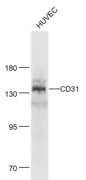 Sample: Sample:Huvec(Human) Cell Lysate at 30 ug Primary: Anti-CD31 (bs-0468R) at 1/1000 dilution Secondary: IRDye800CW Goat Anti-Rabbit IgG at 1/20000 dilution Predicted band size: 78 kD Observed band size: 140 kD 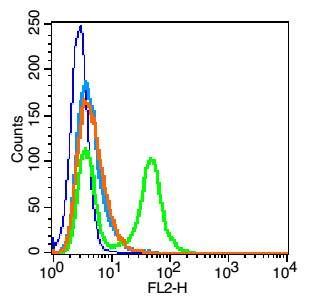 Blank control: HUVEC cells(blue). Blank control: HUVEC cells(blue).Primary Antibody:Rabbit Anti-CD31 antibody(bs-0468R), Dilution: 1μg in 100 μL 1X PBS containing 0.5% BSA; Isotype Control Antibody: Rabbit IgG(orange) ,used under the same conditions ); Secondary Antibody: Goat anti-rabbit IgG-PE(white blue), Dilution: 1:200 in 1 X PBS containing 0.5% BSA. Protocol The cells were fixed with 2% paraformaldehyde (10 min) .Primary antibody (bs-0468R, 1μg /1x10^6 cells) were incubated for 30 min on the ice, followed by 1 X PBS containing 0.5% BSA + 1 0% goat serum (15 min) to block non-specific protein-protein interactions. Then the Goat Anti-rabbit IgG/PE antibody was added into the blocking buffer mentioned above to react with the primary antibody at 1/200 dilution for 30 min on ice. Acquisition of 20,000 events was performed. 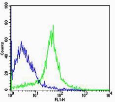 Cell:R.spleen Cell:R.spleenConcentration:1:100 Host/Isotype:Rabbit/IgG Flow cytometric analysis of Rabbit IgG isotype control (Cat#: bs-0468R) on R.spleen(green) compared with control in the absence of primary antibody (blue) followed by Alexa Fluor 488-conjugated goat anti-rabbit IgG(H+L) secondary antibody . |


