
上海细胞库
人源细胞系| 稳转细胞系| 基因敲除株| 基因点突变细胞株| 基因过表达细胞株| 重组细胞系| 猪的细胞系| 马细胞系| 兔的细胞系| 犬的细胞系| 山羊的细胞系| 鱼的细胞系| 猴的细胞系| 仓鼠的细胞系| 狗的细胞系| 牛的细胞| 大鼠细胞系| 小鼠细胞系| 其他细胞系|
人源细胞系| 稳转细胞系| 基因敲除株| 基因点突变细胞株| 基因过表达细胞株| 重组细胞系| 猪的细胞系| 马细胞系| 兔的细胞系| 犬的细胞系| 山羊的细胞系| 鱼的细胞系| 猴的细胞系| 仓鼠的细胞系| 狗的细胞系| 牛的细胞| 大鼠细胞系| 小鼠细胞系| 其他细胞系|

| 规格 | 价格 | 库存 |
|---|---|---|
| 50ul | ¥ 1280 | 200 |
| 100ul | ¥ 1980 | 200 |
| 200ul | ¥ 2980 | 200 |
| 中文名称 | 半胱氨酸蛋白酶8抗体 |
| 别 名 | Caspase-8 p10; Caspase-8 subunit p10; ALPS2B; Amyotrophic lateral sclerosis 2 chromosomal region candidate gene 12 protein; Apoptosis related cysteine peptidase; Apoptotic cysteine protease; Apoptotic protease Mch 5; Apoptotic protease Mch-5; Apoptotic protease Mch5; CAP 4; CAP4; CASP 8; CASP-8; Caspase-8; Caspase8; CASP8; CASP8_HUMAN; Caspase8; Caspase 8 apoptosis related cysteine peptidase; Caspase8; CED 3; FADD homologous ICE/CED 3 like protease; FADD Homologous ICE/CED3 Like Protease; FADD Like ICE antibody FADD-homologous ICE/CED-3-like protease; FADD-like ICE; FLICE; FLJ17672; ICE like apoptotic protease 5; ICE-like apoptotic protease 5; MACH alpha 1/2/3 protein; MACH antibody MACH beta 1/2/3/4 protein antibody MCH 5; MCH5 |
| 研究领域 | 细胞生物 神经生物学 细胞凋亡 |
| 抗体来源 | Rabbit |
| 克隆类型 | Polyclonal |
| 交叉反应 | Human, Rat, Sheep, (predicted: Mouse, Chicken, Dog, Pig, Cow, ) |
| 产品应用 | ELISA=1:500-1000 IHC-P=1:100-500 IHC-F=1:100-500 Flow-Cyt=1μg/Test ICC=1:100 IF=1:100-500 (石蜡切片需做抗原修复) not yet tested in other applications. optimal dilutions/concentrations should be determined by the end user. |
| 分 子 量 | 12/55kDa |
| 细胞定位 | 细胞浆 |
| 性 状 | Liquid |
| 浓 度 | 1mg/ml |
| 免 疫 原 | KLH conjugated synthetic peptide derived from rat Caspase-8 subunit p10:411-482/482 |
| 亚 型 | IgG |
| 纯化方法 | affinity purified by Protein A |
| 储 存 液 | 0.01M TBS(pH7.4) with 1% BSA, 0.03% Proclin300 and 50% Glycerol. |
| 保存条件 | Shipped at 4℃. Store at -20 °C for one year. Avoid repeated freeze/thaw cycles. |
| PubMed | PubMed |
| 产品介绍 | Caspases are cysteine proteases, expressed as inactive precursors, that mediate apoptosis by proteolysis of specific substrates. Caspases have the ability to cleave after aspartic acid residues. There are two classes of caspases involved in apoptosis; initiators (activation by receptor cluster) and effectors (activation by mitochondrial permeability transition). Proapoptotic signals autocatalytically activate initiator caspases, such as Caspase 8 and Caspase 9. Activated initiator caspases then process effector caspases, such as Caspase 3 and Caspase 7, which in turn cause cell collapse. Function: Most upstream protease of the activation cascade of caspases responsible for the TNFRSF6/FAS mediated and TNFRSF1A induced cell death. Binding to the adapter molecule FADD recruits it to either receptor. The resulting aggregate called death-inducing signaling complex (DISC) performs CASP8 proteolytic activation. The active dimeric enzyme is then liberated from the DISC and free to activate downstream apoptotic proteases. Proteolytic fragments of the N-terminal propeptide (termed CAP3, CAP5 and CAP6) are likely retained in the DISC. Cleaves and activates CASP3, CASP4, CASP6, CASP7, CASP9 and CASP10. May participate in the GZMB apoptotic pathways. Cleaves ADPRT. Hydrolyzes the small-molecule substrate, Ac-Asp-Glu-Val-Asp-|-AMC. Likely target for the cowpox virus CRMA death inhibitory protein. Isoform 5, isoform 6, isoform 7 and isoform 8 lack the catalytic site and may interfere with the pro-apoptotic activity of the complex. Subunit: Heterotetramer that consists of two anti-parallel arranged heterodimers, each one formed by a 18 kDa (p18) and a 10 kDa (p10) subunit. Interacts with FADD, CFLAR and PEA15. Isoform 9 interacts at the endoplasmic reticulum with a complex containing BCAP31, BAP29, BCL2 and/or BCL2L1. Interacts with TNFAIP8L2. Subcellular Location: Cytoplasm. Tissue Specificity: Isoform 1, isoform 5 and isoform 7 are expressed in a wide variety of tissues. Highest expression in peripheral blood leukocytes, spleen, thymus and liver. Barely detectable in brain, testis and skeletal muscle. DISEASE: Defects in CASP8 are the cause of caspase-8 deficiency (CASP8D) [MIM:607271]. CASP8D is a disorder resembling autoimmune lymphoproliferative syndrome (ALPS). It is characterized by lymphadenopathy, splenomegaly, and defective CD95-induced apoptosis of peripheral blood lymphocytes (PBLs). It leads to defects in activation of T-lymphocytes, B-lymphocytes, and natural killer cells leading to immunodeficiency characterized by recurrent sinopulmonary and herpes simplex virus infections and poor responses to immunization. Similarity: Belongs to the peptidase C14A family. Contains 2 DED (death effector) domains. SWISS: O89110 Gene ID: 841 Database links: Entrez Gene: 841 Human Entrez Gene: 12370 Mouse Entrez Gene: 54474 Rat Entrez Gene: 64044 Rat Omim: 601763 Human SwissProt: Q14790 Human SwissProt: O89110 Mouse SwissProt: Q9JHX4 Rat Unigene: 599762 Human Unigene: 655983 Human Unigene: 336851 Mouse Important Note: This product as supplied is intended for research use only, not for use in human, therapeutic or diagnostic applications. |
| 产品图片 | 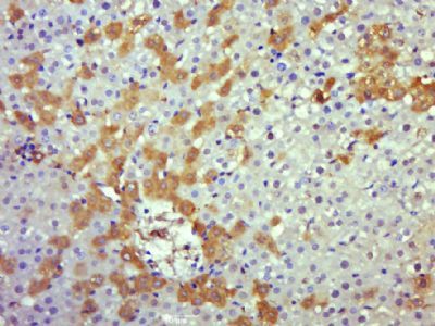 Paraformaldehyde-fixed, paraffin embedded (rat liver); Antigen retrieval by boiling in sodium citrate buffer (pH6.0) for 15min; Block endogenous peroxidase by 3% hydrogen peroxide for 20 minutes; Blocking buffer (normal goat serum) at 37°C for 30min; Antibody incubation with (Caspase 8) Polyclonal Antibody, Unconjugated (bs-0052R) at 1:500 overnight at 4°C, followed by operating according to SP Kit(Rabbit) (sp-0023) instructionsand DAB staining. Paraformaldehyde-fixed, paraffin embedded (rat liver); Antigen retrieval by boiling in sodium citrate buffer (pH6.0) for 15min; Block endogenous peroxidase by 3% hydrogen peroxide for 20 minutes; Blocking buffer (normal goat serum) at 37°C for 30min; Antibody incubation with (Caspase 8) Polyclonal Antibody, Unconjugated (bs-0052R) at 1:500 overnight at 4°C, followed by operating according to SP Kit(Rabbit) (sp-0023) instructionsand DAB staining.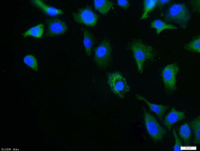 Tissue/cell: Hela cell; 4% Paraformaldehyde-fixed; Triton X-100 at room temperature for 20 min; Blocking buffer (normal goat serum, C-0005) at 37°C for 20 min; Antibody incubation with (Caspase 8) polyclonal Antibody, Unconjugated (bs-0052R) 1:100, 90 minutes at 37°C; followed by a FITC conjugated Goat Anti-Rabbit IgG antibody at 37°C for 90 minutes, DAPI (blue, C02-04002) was used to stain the cell nuclei. Tissue/cell: Hela cell; 4% Paraformaldehyde-fixed; Triton X-100 at room temperature for 20 min; Blocking buffer (normal goat serum, C-0005) at 37°C for 20 min; Antibody incubation with (Caspase 8) polyclonal Antibody, Unconjugated (bs-0052R) 1:100, 90 minutes at 37°C; followed by a FITC conjugated Goat Anti-Rabbit IgG antibody at 37°C for 90 minutes, DAPI (blue, C02-04002) was used to stain the cell nuclei.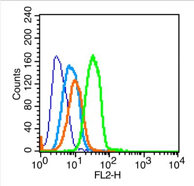 Blank control (blue line): U251 (blue). Blank control (blue line): U251 (blue).Primary Antibody (green line): Rabbit Anti-caspase-8 antibody (bs-0052R) Dilution: 1μg /10^6 cells; Isotype Control Antibody (orange line): Rabbit IgG . Secondary Antibody (white blue line): Goat anti-rabbit IgG-PE Dilution: 1μg /test. Protocol The cells were fixed with 70% ethanol overnight at 4℃ and then permeabilized with 0.1% PBS-Tween for 20 min at room temperature. Cells stained with Primary Antibody for 30 min at room temperature. The cells were then incubated in 1 X PBS/2%BSA/10% goat serum to block non-specific protein-protein interactions followed by the antibody for 15 min at room temperature. The secondary antibody used for 40 min at room temperature. Acquisition of 20,000 events was performed. |


