
上海细胞库
人源细胞系| 稳转细胞系| 基因敲除株| 基因点突变细胞株| 基因过表达细胞株| 重组细胞系| 猪的细胞系| 马细胞系| 兔的细胞系| 犬的细胞系| 山羊的细胞系| 鱼的细胞系| 猴的细胞系| 仓鼠的细胞系| 狗的细胞系| 牛的细胞| 大鼠细胞系| 小鼠细胞系| 其他细胞系|
人源细胞系| 稳转细胞系| 基因敲除株| 基因点突变细胞株| 基因过表达细胞株| 重组细胞系| 猪的细胞系| 马细胞系| 兔的细胞系| 犬的细胞系| 山羊的细胞系| 鱼的细胞系| 猴的细胞系| 仓鼠的细胞系| 狗的细胞系| 牛的细胞| 大鼠细胞系| 小鼠细胞系| 其他细胞系|

| 规格 | 价格 | 库存 |
|---|---|---|
| 50ul | ¥ 980 | 200 |
| 100ul | ¥ 1680 | 200 |
| 200ul | ¥ 2480 | 200 |
| 中文名称 | 血管紧张素Ⅱ受体1抗体 |
| 别 名 | AG2S; Agtr 1; Agtr1; AGTR1_HUMAN; Agtr1a; AGTR1B; Ang II; Angiotensin II receptor type 1; Angiotensin II type 1 receptor; Angiotensin II type-1 receptor; Angiotensin receptor 1; Angiotensin receptor 1B; AT 1B; AT 1r; AT-1B; AT-1r; AT1; At1a; AT1AR; AT1B; AT1BR; AT1R; AT2R1; AT2R1A; AT2R1B; HAT1R; Type 1 angiotensin II receptor; Type 1B angiotensin II receptor; Type-1 angiotensin II receptor. |
| 研究领域 | 肿瘤 心血管 细胞生物 免疫学 血管内皮细胞 |
| 抗体来源 | Rabbit |
| 克隆类型 | Polyclonal |
| 交叉反应 | Mouse, Rat, (predicted: Human, ) |
| 产品应用 | WB=1:500-2000 ELISA=1:500-1000 IHC-P=1:100-500 IHC-F=1:100-500 IF=1:100-500 (石蜡切片需做抗原修复) not yet tested in other applications. optimal dilutions/concentrations should be determined by the end user. |
| 分 子 量 | 41kDa |
| 细胞定位 | 细胞膜 |
| 性 状 | Liquid |
| 浓 度 | 1mg/ml |
| 免 疫 原 | KLH conjugated synthetic peptide derived from human AT2R:151-250/359 |
| 亚 型 | IgG |
| 纯化方法 | affinity purified by Protein A |
| 储 存 液 | 0.01M TBS(pH7.4) with 1% BSA, 0.03% Proclin300 and 50% Glycerol. |
| 保存条件 | Shipped at 4℃. Store at -20 °C for one year. Avoid repeated freeze/thaw cycles. |
| PubMed | PubMed |
| 产品介绍 | Angiotensin II (Ang II) is an important physiological effector of blood pressure and volume regulation through vasoconstriction, aldosterone release, sodium uptake and thirst stimulation. Although Ang II interacts with two types of cell surface receptors, AT1 and AT2, most of the major cardiovascular effects seem to be mediated through AT1. Molecular cloning of the AT1 protein has shown it to be a member of the G protein-associated seven transmembrane protein receptor family. Ang II treatment of cells results in activation of several signal transduction pathways as evidenced by tyrosine phosphorylation of several proteins and induction of others. PLC?is phosphorylated after 30 seconds of treatment with Angiotensin II, indicating this as an early signal transduction event. Ang II treatment also stimulates phosphorylation of Shc, FAK and MAP kinases, and induces MKP-1, indicating stimulation of growth factor pathways. Ang II stimulation through AT1 has been shown to activate the JAK/Stat pathway involving a direct interaction between JAK2 and AT1 as demonstrated by coimmunoprecipitation. The AT1 receptor has no cytoplasmic kinase domain, but is able to function as a substrate for Src kinases and has several putative phosphorylation sites. Function: Receptor for angiotensin II. Mediates its action by association with G proteins that activate a phosphatidylinositol-calcium second messenger system. Subunit: Interacts with MAS1 (Probable). Interacts with ARRB1 (By similarity). Subcellular Location: Cell membrane; Multi-pass membrane protein. Tissue Specificity: Liver, lung, adrenal and adrenocortical adenomas. Post-translational modifications: C-terminal Ser or Thr residues may be phosphorylated. DISEASE: Defects in AGTR1 are a cause of renal tubular dysgenesis (RTD). RTD is an autosomal recessive severe disorder of renal tubular development characterized by persistent fetal anuria and perinatal death, probably due to pulmonary hypoplasia from early-onset oligohydramnios (the Potter phenotype). Similarity: Belongs to the G-protein coupled receptor 1 family. SWISS: P30556 Gene ID: 185 Database links: Entrez Gene: 185 Human Entrez Gene: 11607 Mouse Entrez Gene: 11608 Mouse Entrez Gene: 24180 Rat Entrez Gene: 81638 Rat Omim: 106165 Human SwissProt: P30556 Human SwissProt: P29754 Mouse SwissProt: P29755 Mouse SwissProt: P25095 Rat SwissProt: P29089 Rat Unigene: 477887 Human Unigene: 728754 Human Important Note: This product as supplied is intended for research use only, not for use in human, therapeutic or diagnostic applications. 主要用于心、脑、血管病及肿瘤的基础研究。 |
| 产品图片 | 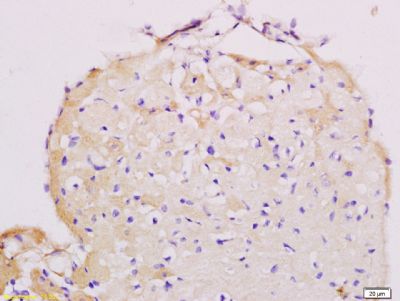 Tissue/cell: mouse heart tissue; 4% Paraformaldehyde-fixed and paraffin-embedded; Tissue/cell: mouse heart tissue; 4% Paraformaldehyde-fixed and paraffin-embedded;Antigen retrieval: citrate buffer ( 0.01M, pH 6.0 ), Boiling bathing for 15min; Block endogenous peroxidase by 3% Hydrogen peroxide for 30min; Blocking buffer (normal goat serum,C-0005) at 37℃ for 20 min; Incubation: Anti-AT2R/AT1 Polyclonal Antibody, Unconjugated(bs-0438R) 1:200, overnight at 4°C, followed by conjugation to the secondary antibody(SP-0023) and DAB(C-0010) staining 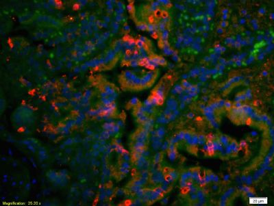 Tissue/cell: rat colon tissue;4% Paraformaldehyde-fixed and paraffin-embedded; Tissue/cell: rat colon tissue;4% Paraformaldehyde-fixed and paraffin-embedded;Antigen retrieval: citrate buffer ( 0.01M, pH 6.0 ), Boiling bathing for 15min; Blocking buffer (normal goat serum,C-0005) at 37℃ for 20 min; Incubation: Anti-AT2R/AT1 Polyclonal Antibody, Unconjugated(bs-0438R) 1:200, overnight at 4°C; The secondary antibody was Goat Anti-Rabbit IgG, Cy3 conjugated(bs-0295G-Cy3)used at 1:200 dilution for 40 minutes at 37°C. DAPI(5ug/ml,blue,C-0033) was used to stain the cell nuclei 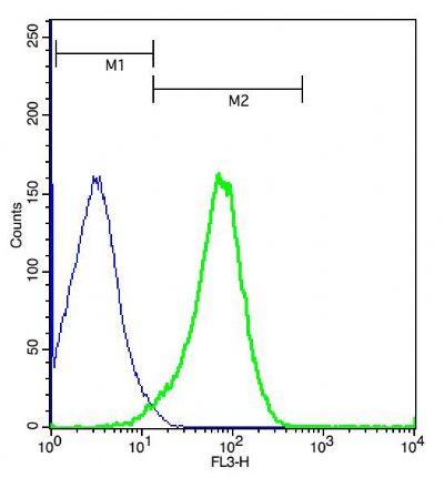 Cell: (mo)Splenocytes( 2% Paraformaldehyde fixed for 10 minutes). Cell: (mo)Splenocytes( 2% Paraformaldehyde fixed for 10 minutes).Concentration:1:50. Incubation: 40 minutes. Host/Blank: (mo)Splenocytes. Flow cytometric analysis of Rabbit Anti-AT2R/PE-Cy5.5 Conjugated antibody (bs-0438R-PE-Cy5.5)(green) compared with control in the absence of primary antibody (blue) followed by mouse spleen cells. |


