
上海细胞库
人源细胞系| 稳转细胞系| 基因敲除株| 基因点突变细胞株| 基因过表达细胞株| 重组细胞系| 猪的细胞系| 马细胞系| 兔的细胞系| 犬的细胞系| 山羊的细胞系| 鱼的细胞系| 猴的细胞系| 仓鼠的细胞系| 狗的细胞系| 牛的细胞| 大鼠细胞系| 小鼠细胞系| 其他细胞系|
人源细胞系| 稳转细胞系| 基因敲除株| 基因点突变细胞株| 基因过表达细胞株| 重组细胞系| 猪的细胞系| 马细胞系| 兔的细胞系| 犬的细胞系| 山羊的细胞系| 鱼的细胞系| 猴的细胞系| 仓鼠的细胞系| 狗的细胞系| 牛的细胞| 大鼠细胞系| 小鼠细胞系| 其他细胞系|

| 规格 | 价格 | 库存 |
|---|---|---|
| 50ul | ¥ 980 | 200 |
| 100ul | ¥ 1680 | 200 |
| 200ul | ¥ 2480 | 200 |
| 中文名称 | 自噬蛋白5/细胞凋亡的特异性蛋白抗体 |
| 别 名 | ATG5; APG 5; APG 5L; APG5; APG5 autophagy 5 like; APG5 like; APG5-like; APG5L; Apoptosis specific protein; Apoptosis-specific protein; ASP; ATG 5; ATG5; ATG5 autophagy related 5 homolog; Autophagy protein 5; hAPG5; Homolog of S Cerevisiae autophagy 5; ATG5_HUMAN. |
| 研究领域 | 肿瘤 细胞生物 免疫学 细胞凋亡 细胞自噬 |
| 抗体来源 | Rabbit |
| 克隆类型 | Polyclonal |
| 交叉反应 | Human, Mouse, (predicted: Rat, Chicken, Dog, Pig, Cow, Horse, Rabbit, ) |
| 产品应用 | WB=1:500-2000 ELISA=1:500-1000 IHC-P=1:100-500 IHC-F=1:100-500 Flow-Cyt=1ug/Test IF=1:100-500 (石蜡切片需做抗原修复) not yet tested in other applications. optimal dilutions/concentrations should be determined by the end user. |
| 分 子 量 | 32kDa |
| 细胞定位 | 细胞浆 |
| 性 状 | Liquid |
| 浓 度 | 1mg/ml |
| 免 疫 原 | KLH conjugated synthetic peptide derived from human APG5L:101-200/275 |
| 亚 型 | IgG |
| 纯化方法 | affinity purified by Protein A |
| 储 存 液 | 0.01M TBS(pH7.4) with 1% BSA, 0.03% Proclin300 and 50% Glycerol. |
| 保存条件 | Shipped at 4℃. Store at -20 °C for one year. Avoid repeated freeze/thaw cycles. |
| PubMed | PubMed |
| 产品介绍 | In yeast, autophagy is an essential process for survival during nutrient starvation and cell differentiation. The process of autophagy is characterized as a non-selective degradation of cytoplasmic proteins into membrane stuctures called autophagosomes, and it is dependent on several proteins, including the autophagy proteins APG5 and APG7. Yeast Apg7 and the human homolog, APG7, share similarities with the ubiquitin-activating enzyme E1 in Saccharomyces cerevisiae and are likewise responsible for enzymatically activating the autophagy conjugation system. Apg5 and the human homolog, APG5 (also designated apoptosis-specific protein or APS), function as substrates for the autophagy protein Apg12. These proteins are covalently bonded together to form Apg12/APG5 conjugates, which are required for the progression of autophagy. Function: Required for autophagy. Conjugates to ATG12 and associates with isolation membrane to form cup-shaped isolation membrane and autophagosome. The conjugate detaches from the membrane immediately before or after autophagosome formation is completed (By similarity). May play an important role in the apoptotic process, possibly within the modified cytoskeleton. Its expression is a relatively late event in the apoptotic process, occurring downstream of caspase activity. Subunit: The ATG5-ATG12 conjugate forms a complex with several units of ATG16. Interacts with TECPR1; the interaction is direct and does not take place when ATG16 is associated with the ATG5-ATG12 conjugate. Subcellular Location: Cytoplasm. Note=Colocalizes with nonmuscle actin. Tissue Specificity: Ubiquitous. The mRNA is present at similar levels in viable and apoptotic cells, whereas the protein is dramatically highly expressed in apoptotic cells. Post-translational modifications: Conjugated to ATG12; which is essential for autophagy, but is not required for association with isolation membrane. Similarity: Belongs to the ATG5 family. SWISS: Q9H1Y0 Gene ID: 9474 Database links: Entrez Gene: 9474 Human Entrez Gene: 11793 Mouse Entrez Gene: 365601 Rat Omim: 604261 Human SwissProt: Q9H1Y0 Human SwissProt: Q99J83 Mouse SwissProt: Q3MQ06 Rat Unigene: 486063 Human Unigene: 22264 Mouse Unigene: 98385 Rat Important Note: This product as supplied is intended for research use only, not for use in human, therapeutic or diagnostic applications. |
| 产品图片 | 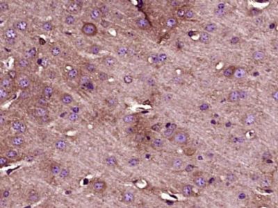 Paraformaldehyde-fixed, paraffin embedded (Mouse brain); Antigen retrieval by boiling in sodium citrate buffer (pH6.0) for 15min; Block endogenous peroxidase by 3% hydrogen peroxide for 20 minutes; Blocking buffer (normal goat serum) at 37°C for 30min; Antibody incubation with (ATG5) Polyclonal Antibody, Unconjugated (bs-4005R) at 1:400 overnight at 4°C, followed by operating according to SP Kit(Rabbit) (sp-0023) instructionsand DAB staining. Paraformaldehyde-fixed, paraffin embedded (Mouse brain); Antigen retrieval by boiling in sodium citrate buffer (pH6.0) for 15min; Block endogenous peroxidase by 3% hydrogen peroxide for 20 minutes; Blocking buffer (normal goat serum) at 37°C for 30min; Antibody incubation with (ATG5) Polyclonal Antibody, Unconjugated (bs-4005R) at 1:400 overnight at 4°C, followed by operating according to SP Kit(Rabbit) (sp-0023) instructionsand DAB staining.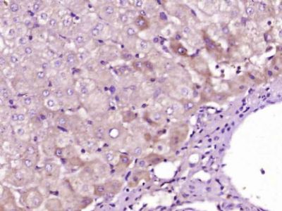 Paraformaldehyde-fixed, paraffin embedded (Human liver carcinoma); Antigen retrieval by boiling in sodium citrate buffer (pH6.0) for 15min; Block endogenous peroxidase by 3% hydrogen peroxide for 20 minutes; Blocking buffer (normal goat serum) at 37°C for 30min; Antibody incubation with (ATG5) Polyclonal Antibody, Unconjugated (bs-4005R) at 1:400 overnight at 4°C, followed by operating according to SP Kit(Rabbit) (sp-0023) instructionsand DAB staining. Paraformaldehyde-fixed, paraffin embedded (Human liver carcinoma); Antigen retrieval by boiling in sodium citrate buffer (pH6.0) for 15min; Block endogenous peroxidase by 3% hydrogen peroxide for 20 minutes; Blocking buffer (normal goat serum) at 37°C for 30min; Antibody incubation with (ATG5) Polyclonal Antibody, Unconjugated (bs-4005R) at 1:400 overnight at 4°C, followed by operating according to SP Kit(Rabbit) (sp-0023) instructionsand DAB staining.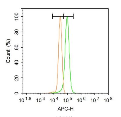 Blank control: A431. Blank control: A431.Primary Antibody (green line): Rabbit Anti-ATG5 antibody (bs-4005R) Dilution: 1μg /10^6 cells; Isotype Control Antibody (orange line): Rabbit IgG . Secondary Antibody: Goat anti-rabbit IgG-AF647 Dilution: 1μg /test. Protocol The cells were fixed with 4% PFA (10min at room temperature)and then permeabilized with 0.1% PBST for 20 min at room temperature. The cells were then incubated in 5%BSA to block non-specific protein-protein interactions for 30 min at room temperature .Cells stained with Primary Antibody for 30 min at room temperature. The secondary antibody used for 40 min at room temperature. Acquisition of 20,000 events was performed. |


