
上海细胞库
人源细胞系| 稳转细胞系| 基因敲除株| 基因点突变细胞株| 基因过表达细胞株| 重组细胞系| 猪的细胞系| 马细胞系| 兔的细胞系| 犬的细胞系| 山羊的细胞系| 鱼的细胞系| 猴的细胞系| 仓鼠的细胞系| 狗的细胞系| 牛的细胞| 大鼠细胞系| 小鼠细胞系| 其他细胞系|
人源细胞系| 稳转细胞系| 基因敲除株| 基因点突变细胞株| 基因过表达细胞株| 重组细胞系| 猪的细胞系| 马细胞系| 兔的细胞系| 犬的细胞系| 山羊的细胞系| 鱼的细胞系| 猴的细胞系| 仓鼠的细胞系| 狗的细胞系| 牛的细胞| 大鼠细胞系| 小鼠细胞系| 其他细胞系|

| 规格 | 价格 | 库存 |
|---|---|---|
| 50ul | ¥ 980 | 200 |
| 100ul | ¥ 1680 | 200 |
| 200ul | ¥ 2480 | 200 |
| 中文名称 | 生长分化因子9抗体 |
| 别 名 | GDF9_HUMAN; GDF9; GDF-9; GDF 9; Growth differentiation factor 9. |
| 研究领域 | 心血管 免疫学 信号转导 干细胞 生长因子和激素 细胞分化 |
| 抗体来源 | Rabbit |
| 克隆类型 | Polyclonal |
| 交叉反应 | Human, Mouse, Rat, (predicted: Chicken, Dog, Pig, Cow, Horse, Sheep, ) |
| 产品应用 | WB=1:500-2000 ELISA=1:500-1000 IHC-P=1:100-500 Flow-Cyt=1ug/Test (石蜡切片需做抗原修复) not yet tested in other applications. optimal dilutions/concentrations should be determined by the end user. |
| 分 子 量 | 15kDa |
| 细胞定位 | 分泌型蛋白 |
| 性 状 | Liquid |
| 浓 度 | 1mg/ml |
| 免 疫 原 | KLH conjugated synthetic peptide derived from human GDF9:301-400/454 |
| 亚 型 | IgG |
| 纯化方法 | affinity purified by Protein A |
| 储 存 液 | 0.01M TBS(pH7.4) with 1% BSA, 0.03% Proclin300 and 50% Glycerol. |
| 保存条件 | Shipped at 4℃. Store at -20 °C for one year. Avoid repeated freeze/thaw cycles. |
| PubMed | PubMed |
| 产品介绍 | GDF 9 is a member of the bone morphogenetic protein (BMP) family and the TGF-beta superfamily. This group of proteins is characterized by a polybasic proteolytic processing site which is cleaved to produce a mature protein containing seven conserved cysteine residues. The members of this family are regulators of cell synthesized by ovarian somatic cells directly affect oocyte growth and function. GDF 9 is expressed in oocytes and is thought to be required for ovarian folliculogenesis. Function: Required for ovarian folliculogenesis. Promotes primordial follicle development. Stimulates granulosa cell proliferation. Promotes cell transition from G0/G1 to S and G2/M phases, through an increase of CCND1 and CCNE1 expression, and RB1 phosphorylation. It regulates STAR expression and cAMP-dependent progesterone release in granulosa and thecal cells. Attenuates the suppressive effects of activin A on STAR expression and progesterone production by increasing the expression of inhibin B. It suppresses FST and FSTL3 production in granulosa-lutein cells. Subunit: Homodimer or heterodimer (Potential). But, in contrast to other members of this family, cannot be disulfide-linked (By similarity). Subcellular Location: Secreted (By similarity). Tissue Specificity: Expressed in ovarian granulosa cells. Present in oocytes of primary follicles (at protein level). Post-translational modifications: Phosphorylated; phosphorylation is critical for GDF9 function. In vitro, can be phosphorylated by CK at Ser-325. DISEASE: Note=Altered GDF9 function may be involved in ovarian disorders. Rare variants in GDF9 have been found in patients with premature ovarian failure and mothers of dizygotic twins. Similarity: Belongs to the TGF-beta family. SWISS: O60383 Gene ID: 2661 Database links: Entrez Gene: 2661 Human Entrez Gene: 14566 Mouse Entrez Gene: 59304 Rat Omim: 601918 Human SwissProt: O60383 Human SwissProt: Q07105 Mouse Unigene: 25022 Human Unigene: 490461 Mouse Unigene: 9714 Mouse Unigene: 42874 Rat Important Note: This product as supplied is intended for research use only, not for use in human, therapeutic or diagnostic applications. GDF9属于转移生长因子–β(TGF-β)及骨形态发生蛋白(BMP)家族成员。 |
| 产品图片 | 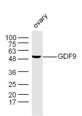 Sample: Ovary (Mouse) Lysate at 40 ug Sample: Ovary (Mouse) Lysate at 40 ugPrimary: Anti-GDF9 (bs-4720R) at 1/300 dilution Secondary: IRDye800CW Goat Anti-Rabbit IgG at 1/20000 dilution Predicted band size: 15 kD Observed band size: 50 kD 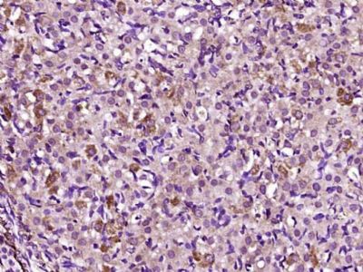 Paraformaldehyde-fixed, paraffin embedded (Rat ovarian); Antigen retrieval by boiling in sodium citrate buffer (pH6.0) for 15min; Block endogenous peroxidase by 3% hydrogen peroxide for 20 minutes; Blocking buffer (normal goat serum) at 37°C for 30min; Antibody incubation with (GDF9) Polyclonal Antibody, Unconjugated (bs-4720R) at 1:400 overnight at 4°C, followed by operating according to SP Kit(Rabbit) (sp-0023) instructionsand DAB staining. Paraformaldehyde-fixed, paraffin embedded (Rat ovarian); Antigen retrieval by boiling in sodium citrate buffer (pH6.0) for 15min; Block endogenous peroxidase by 3% hydrogen peroxide for 20 minutes; Blocking buffer (normal goat serum) at 37°C for 30min; Antibody incubation with (GDF9) Polyclonal Antibody, Unconjugated (bs-4720R) at 1:400 overnight at 4°C, followed by operating according to SP Kit(Rabbit) (sp-0023) instructionsand DAB staining.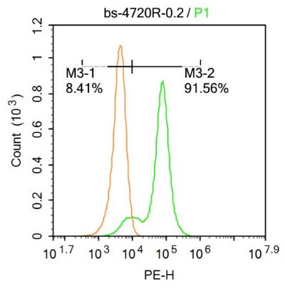 Blank control:Molt-4. Blank control:Molt-4.Primary Antibody (green line): Rabbit Anti-GDF9 antibody (bs-4720) Dilution: 0.2μg /10^6 cells; Isotype Control Antibody (orange line): Rabbit IgG . Secondary Antibody : Goat anti-rabbit IgG-PE Dilution: 0.2μg /test. Protocol The cells were fixed with 4% PFA (10min at room temperature)and then permeabilized with 0.1% PBST for 20 min at room temperature. The cells were then incubated in 5%BSA to block non-specific protein-protein interactions for 30 min at at room temperature .Cells stained with Primary Antibody for 30 min at room temperature. The secondary antibody used for 40 min at room temperature. Acquisition of 20,000 events was performed. 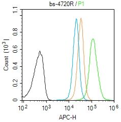 Blank control (Black line): Molt4 (Black). Blank control (Black line): Molt4 (Black).Primary Antibody (green line):Rabbit Anti-GDF9 antibody (bs-4720R) Dilution: 3μg /10^6 cells; Isotype Control Antibody (orange line): Rabbit IgG . Secondary Antibody (white blue line): Goat anti-rabbit IgG-AF647 Dilution: 3μg /test. Protocol The cells were fixed with 4% PFA (10min at room temperature)and then permeabilized with PBST for 20 min at room temperature. The cells were then incubated in 5%BSA to block non-specific protein-protein interactions for 30 min at room temperature .Cells stained with Primary Antibody for 30 min at room temperature. The secondary antibody used for 40 min at room temperature. Acquisition of 20,000 events was performed. |


