
上海细胞库
人源细胞系| 稳转细胞系| 基因敲除株| 基因点突变细胞株| 基因过表达细胞株| 重组细胞系| 猪的细胞系| 马细胞系| 兔的细胞系| 犬的细胞系| 山羊的细胞系| 鱼的细胞系| 猴的细胞系| 仓鼠的细胞系| 狗的细胞系| 牛的细胞| 大鼠细胞系| 小鼠细胞系| 其他细胞系|
人源细胞系| 稳转细胞系| 基因敲除株| 基因点突变细胞株| 基因过表达细胞株| 重组细胞系| 猪的细胞系| 马细胞系| 兔的细胞系| 犬的细胞系| 山羊的细胞系| 鱼的细胞系| 猴的细胞系| 仓鼠的细胞系| 狗的细胞系| 牛的细胞| 大鼠细胞系| 小鼠细胞系| 其他细胞系|

| 规格 | 价格 | 库存 |
|---|---|---|
| 100ul | ¥ 1680 | 200 |
| 200ul | ¥ 2480 | 200 |
| 中文名称 | 内皮分化型G蛋白偶联受体3抗体 |
| 别 名 | EDG 3; EDG3; Endothelial differentiation G protein coupled receptor 3; Endothelial differentiation G-protein coupled receptor 3; Endothelial differentiation gene 3; Endothelial differentiation sphingolipid G protein coupled receptor 3; FLJ37523; FLJ93220; G protein coupled receptor endothelial differentiation gene 3; LPB 3; LPB3; S1P receptor 3; S1P receptor Edg 3; S1P receptor Edg-3; S1P receptor EDG3; S1P3; S1PR3; S1PR3_HUMAN; Sphingosine 1 phosphate receptor 3; Sphingosine 1 phosphate receptor Edg 3; Sphingosine 1 phosphate receptor Edg3; Sphingosine 1-phosphate receptor 3; Sphingosine 1-phosphate receptor Edg-3. |
| 研究领域 | 细胞生物 信号转导 转录调节因子 G蛋白偶联受体 G蛋白信号 |
| 抗体来源 | Rabbit |
| 克隆类型 | Polyclonal |
| 交叉反应 | Human, Mouse, Rat, (predicted: Cow, Rabbit, ) |
| 产品应用 | WB=1:500-2000 ELISA=1:500-1000 IHC-P=1:100-500 IHC-F=1:100-500 Flow-Cyt=0.2u/test ICC=1:100-500 IF=1:100-500 (石蜡切片需做抗原修复) not yet tested in other applications. optimal dilutions/concentrations should be determined by the end user. |
| 分 子 量 | 42kDa |
| 细胞定位 | 细胞膜 |
| 性 状 | Liquid |
| 浓 度 | 1mg/ml |
| 免 疫 原 | KLH conjugated synthetic peptide derived from human EDG3:145-250/378 |
| 亚 型 | IgG |
| 纯化方法 | affinity purified by Protein A |
| 储 存 液 | 0.01M TBS(pH7.4) with 1% BSA, 0.03% Proclin300 and 50% Glycerol. |
| 保存条件 | Shipped at 4℃. Store at -20 °C for one year. Avoid repeated freeze/thaw cycles. |
| PubMed | PubMed |
| 产品介绍 | This gene encodes a member of the EDG family of receptors, which are G protein-coupled receptors. This protein has been identified as a functional receptor for sphingosine 1-phosphate and likely contributes to the regulation of angiogenesis and vascular endothelial cell function. [provided by RefSeq, Jul 2008]. Function: Receptor for the lysosphingolipid sphingosine 1-phosphate (S1P). S1P is a bioactive lysophospholipid that elicits diverse physiological effect on most types of cells and tissues. When expressed in rat HTC4 hepatoma cells, is capable of mediating S1P-induced cell proliferation and suppression of apoptosis. Subcellular Location: Cell membrane; Multi-pass membrane protein. Tissue Specificity: Expressed in all tissues, but most abundantly in heart, placenta, kidney, and liver. Similarity: Belongs to the G-protein coupled receptor 1 family. SWISS: Q99500 Gene ID: 1903 Database links: Entrez Gene: 1903 Human Entrez Gene: 13610 Mouse Entrez Gene: 306792 Rat Omim: 601965 Human SwissProt: Q99500 Human SwissProt: Q9Z0U9 Mouse Unigene: 585118 Human Unigene: 136736 Mouse Unigene: 23918 Rat Important Note: This product as supplied is intended for research use only, not for use in human, therapeutic or diagnostic applications. |
| 产品图片 | 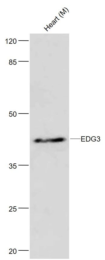 Sample: Sample:Lane1: Heart (Mouse) Lysate at 40 ug Primary: Anti-EDG3 (bs-7541R) at 1/300 dilution Secondary: HRP conjugated Goat-Anti-rabbit IgG (bs-0295G-HRP) at 1/5000 dilution Predicted band size: 42 kD Observed band size: 46 kD 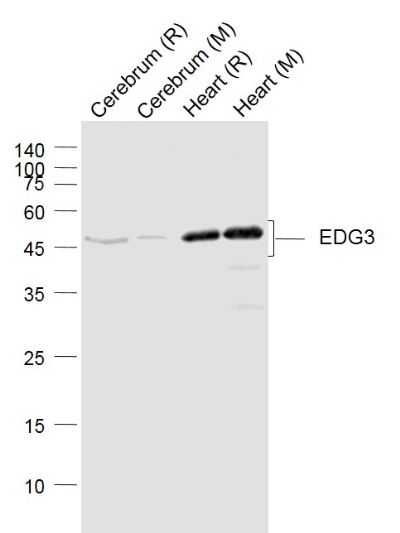 Sample: Sample:Lane 1: Cerebrum (Rat) Lysate at 40 ug Lane 2: Cerebrum (Mouse) Lysate at 40 ug Lane 3: Heart (Rat) Lysate at 40 ug Lane 4: Heart (Mouse) Lysate at 40 ug Primary: Anti-EDG3 (bs-7541R) at 1/1000 dilution Secondary: IRDye800CW Goat Anti-Rabbit IgG at 1/20000 dilution Predicted band size: 46 kD Observed band size: 46 kD 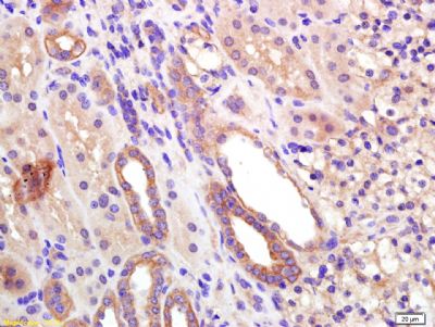 Tissue/cell: human kidney tissue; 4% Paraformaldehyde-fixed and paraffin-embedded; Tissue/cell: human kidney tissue; 4% Paraformaldehyde-fixed and paraffin-embedded;Antigen retrieval: citrate buffer ( 0.01M, pH 6.0 ), Boiling bathing for 15min; Block endogenous peroxidase by 3% Hydrogen peroxide for 30min; Blocking buffer (normal goat serum,C-0005) at 37∩ for 20 min; Incubation: Anti-EDG3 Polyclonal Antibody, Unconjugated(bs-7541R) 1:200, overnight at 4∑C, followed by conjugation to the secondary antibody(SP-0023) and DAB(C-0010) staining 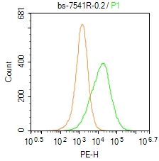 Blank control: Hela. Blank control: Hela.Primary Antibody (green line): Rabbit Anti-EDG3 antibody (bs-7541R) Dilution: 0.2μg /10^6 cells; Isotype Control Antibody (orange line): Rabbit IgG . Secondary Antibody : Goat anti-rabbit IgG-PE Dilution: 1μg /test. Protocol The cells were incubated in 5%BSA to block non-specific protein-protein interactions for 30 min at at room temperature .Cells stained with Primary Antibody for 30 min at room temperature. The secondary antibody used for 40 min at room temperature. Acquisition of 20,000 events was performed. |


