
上海细胞库
人源细胞系| 稳转细胞系| 基因敲除株| 基因点突变细胞株| 基因过表达细胞株| 重组细胞系| 猪的细胞系| 马细胞系| 兔的细胞系| 犬的细胞系| 山羊的细胞系| 鱼的细胞系| 猴的细胞系| 仓鼠的细胞系| 狗的细胞系| 牛的细胞| 大鼠细胞系| 小鼠细胞系| 其他细胞系|
人源细胞系| 稳转细胞系| 基因敲除株| 基因点突变细胞株| 基因过表达细胞株| 重组细胞系| 猪的细胞系| 马细胞系| 兔的细胞系| 犬的细胞系| 山羊的细胞系| 鱼的细胞系| 猴的细胞系| 仓鼠的细胞系| 狗的细胞系| 牛的细胞| 大鼠细胞系| 小鼠细胞系| 其他细胞系|

| 规格 | 价格 | 库存 |
|---|---|---|
| 100ul | ¥ 1880 | 200 |
| 200ul | ¥ 2880 | 200 |
| 中文名称 | 受体结合丝氨酸苏氨酸激酶3抗体 |
| 别 名 | Receptor interacting protein 3; Receptor interacting serine threonine kinase 3; Receptor interacting serine/threonine protein kinase 3; Receptor-interacting protein 3; Receptor-interacting serine/threonine-protein kinase 3; RIP 3; RIP like protein kinase 3; RIP-3; RIP-like protein kinase 3; RIPK 3; RIPK3; RIPK3_HUMAN. |
| 研究领域 | 细胞生物 免疫学 信号转导 细胞凋亡 转录调节因子 激酶和磷酸酶 细胞膜受体 |
| 抗体来源 | Rabbit |
| 克隆类型 | Polyclonal |
| 交叉反应 | Human, Rat, (predicted: Mouse, Dog, Pig, Cow, Rabbit, Guinea Pig, ) |
| 产品应用 | WB=1:500-2000 ELISA=1:500-1000 IHC-P=1:100-500 IHC-F=1:100-500 IF=1:100-500 (石蜡切片需做抗原修复) not yet tested in other applications. optimal dilutions/concentrations should be determined by the end user. |
| 分 子 量 | 57kDa |
| 细胞定位 | 细胞浆 细胞膜 |
| 性 状 | Liquid |
| 浓 度 | 1mg/ml |
| 免 疫 原 | KLH conjugated synthetic peptide derived from human RIPK3:101-230/518 |
| 亚 型 | IgG |
| 纯化方法 | affinity purified by Protein A |
| 储 存 液 | 0.01M TBS(pH7.4) with 1% BSA, 0.03% Proclin300 and 50% Glycerol. |
| 保存条件 | Shipped at 4℃. Store at -20 °C for one year. Avoid repeated freeze/thaw cycles. |
| PubMed | PubMed |
| 产品介绍 | Certain serine/threonine protein kinases, such as ASK1, RIP, DAP, and ZIP kinases, are mediators of apoptosis. Receptor interacting proteins including RIP and RIP2/RICK mediate apoptosis induced by TNFR1 and Fas, two prototype members in the death receptor family. A novel member in the RIP kinase family was recently identified and designated RIP3. RIP3 contains an N terminal kinase domain but, unlike RIP or RIP2, lacks the C-terminal death or CARD domain. RIP3 binds to RIP and TNFR1, mediates TNFR1 induced apoptosis, and attenuates RIP and TNFR1 induced NF-kB activation. Over expression of RIP3 induces apoptosis and NF-kB activation. The messenger RNA of RIP3 is expressed in a subset of adult tissues. Function: Essential for programmed necrosis in response to death-inducing TNF-alpha family members. Upon induction of necrosis, RIPK3 interacts with, and phosphorylates RIPK1 to form a necrosis-inducing complex. RIPK3 binds to and enhances the activity of three metabolic enzymes: GLUL, GLUD1, and PYGL. These metabolic enzymes may eventually stimulate the tricarboxylic acid cycle and oxidative phosphorylation, which could result in enhanced ROS production (By similarity). Subunit: Interacts (via RIP homotypic interaction motif) with RIPK1 (via RIP homotypic interaction motif); this interaction induces RIPK1 phosphorylation and formation of a RIPK1-RIPK3 necrosis-inducing complex. Upon TNF-induced necrosis, the RIPK1-RIPK3 dimer further interacts with PGAM5 and MLKL; the formation of this complex leads to PGAM5 phosphorylation and increase in PGAM5 phosphatase activity. Binds TRAF2 and is recruited to the TNFR-1 signaling complex. Interacts with PYGL, GLUL and GLUD1; these interactions result in activation of these metabolic enzymes. Interacts with BIRC2/c-IAP1, BIRC3/c-IAP2 and XIAP/BIRC4. Subcellular Location: Cytoplasm. Cell membrane. Mitochondrion (Potential). Tissue Specificity: Highly expressed in the pancreas. Detected at lower levels in heart, placenta, lung and kidney. Isoform 3 is significantly increased in colon and lung cancers. Post-translational modifications: RIPK1 and RIPK3 undergo reciprocal auto- and trans-phosphorylation. Phosphorylation of Ser-199 plays a role in the necroptotic function of RIPK3. Polyubiquitinated with 'Lys-48' and 'Lys-63'-linked chains by BIRC2/c-IAP1 and BIRC3/c-IAP2, leading to activation of NF-kappa-B. Similarity: Belongs to the protein kinase superfamily. TKL Ser/Thr protein kinase family. Contains 1 protein kinase domain. SWISS: Q9Y572 Gene ID: 11035 Database links: Entrez Gene: 11035 Human Entrez Gene: 56532 Mouse Omim: 605817 Human SwissProt: Q9Y572 Human SwissProt: Q9QZL0 Mouse Unigene: 268551 Human Unigene: 46612 Mouse Important Note: This product as supplied is intended for research use only, not for use in human, therapeutic or diagnostic applications. |
| 产品图片 | 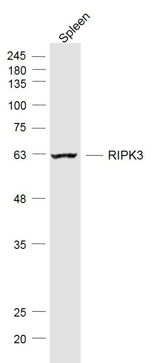 Sample: Sample:Spleen (Mouse) Lysate at 40 ug Primary: Anti-RIPK3 (bs-3551R) at 1/1000 dilution Secondary: IRDye800CW Goat Anti-Rabbit IgG at 1/20000 dilution Predicted band size: 57 kD Observed band size: 57 kD 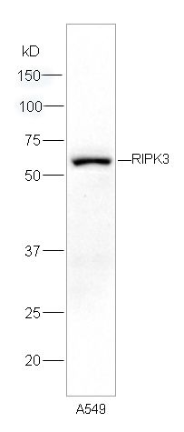 Sample: A549 cells lysates, 30ug; Sample: A549 cells lysates, 30ug;Primary: Anti-RIPK3 (bs-3551R) at 1:300 dilution; Secondary: HRP conjugated Goat Anti-Rabbit IgG(bs-0295G-HRP) at 1: 5000 dilution; Predicted band size : 57kD Observed band size : 57kD 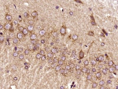 Paraformaldehyde-fixed, paraffin embedded (Rat brain); Antigen retrieval by boiling in sodium citrate buffer (pH6.0) for 15min; Block endogenous peroxidase by 3% hydrogen peroxide for 20 minutes; Blocking buffer (normal goat serum) at 37°C for 30min; Antibody incubation with (RIPK3) Polyclonal Antibody, Unconjugated (bs-3551R) at 1:400 overnight at 4°C, followed by a conjugated secondary antibody (sp-0023) for 20 minutes and DAB staining. Paraformaldehyde-fixed, paraffin embedded (Rat brain); Antigen retrieval by boiling in sodium citrate buffer (pH6.0) for 15min; Block endogenous peroxidase by 3% hydrogen peroxide for 20 minutes; Blocking buffer (normal goat serum) at 37°C for 30min; Antibody incubation with (RIPK3) Polyclonal Antibody, Unconjugated (bs-3551R) at 1:400 overnight at 4°C, followed by a conjugated secondary antibody (sp-0023) for 20 minutes and DAB staining.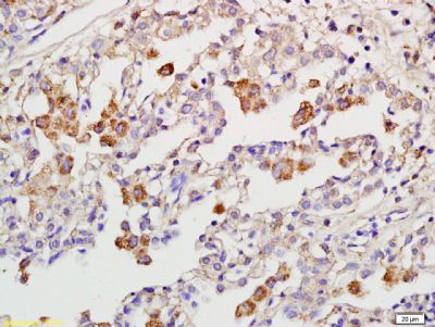 Tissue/cell: human pneumonia tissue; 4% Paraformaldehyde-fixed and paraffin-embedded; Tissue/cell: human pneumonia tissue; 4% Paraformaldehyde-fixed and paraffin-embedded;Antigen retrieval: citrate buffer ( 0.01M, pH 6.0 ), Boiling bathing for 15min; Block endogenous peroxidase by 3% Hydrogen peroxide for 30min; Blocking buffer (normal goat serum,C-0005) at 37℃ for 20 min; Incubation: Anti-RIPK3 Polyclonal Antibody, Unconjugated(bs-3551R) 1:200, overnight at 4°C, followed by conjugation to the secondary antibody(SP-0023) and DAB(C-0010) staining |


