上海细胞库
人源细胞系| 稳转细胞系| 基因敲除株| 基因点突变细胞株| 基因过表达细胞株| 重组细胞系| 猪的细胞系| 马细胞系| 兔的细胞系| 犬的细胞系| 山羊的细胞系| 鱼的细胞系| 猴的细胞系| 仓鼠的细胞系| 狗的细胞系| 牛的细胞| 大鼠细胞系| 小鼠细胞系| 其他细胞系|

| 规格 | 价格 | 库存 |
|---|---|---|
| 50ul | ¥ 980 | 200 |
| 100ul | ¥ 1680 | 200 |
| 200ul | ¥ 2480 | 200 |
| 中文名称 | 细胞色素C抗体 |
| 别 名 | CytC; CYC; CYCS; Cytochrome c somatic; HCS; CYC_HUMAN; Cytochrome-c; MSA06; THC4. |
| 研究领域 | 肿瘤 心血管 细胞生物 神经生物学 信号转导 细胞凋亡 脂蛋白 新陈代谢 线粒体 |
| 抗体来源 | Rabbit |
| 克隆类型 | Polyclonal |
| 交叉反应 | Human, Mouse, Rat, (predicted: Chicken, Pig, Cow, Horse, Rabbit, Guinea Pig, ) |
| 产品应用 | WB=1:500-2000 ELISA=1:500-1000 IHC-P=1:100-500 IHC-F=1:100-500 Flow-Cyt=1μg/Test ICC=1:100-500 IF=1:100-500 (石蜡切片需做抗原修复) not yet tested in other applications. optimal dilutions/concentrations should be determined by the end user. |
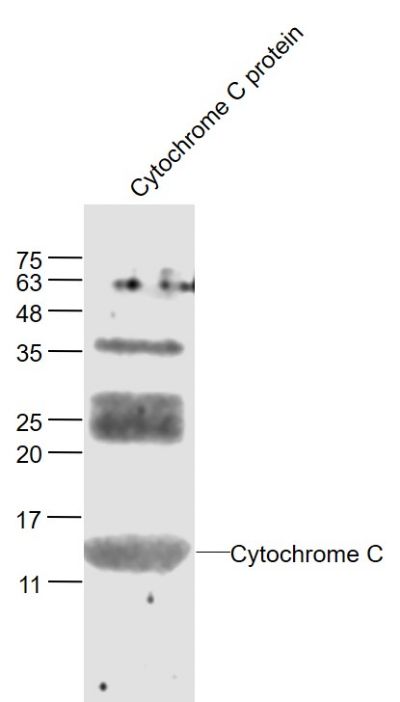
| 分 子 量 | 12.8/26kDa |
| 细胞定位 | 细胞浆 细胞膜 线粒体 |
| 性 状 | Liquid |
| 浓 度 | 1mg/ml |
| 免 疫 原 | KLH conjugated synthetic peptide derived from human Cytochrome C:51-105/105 |
| 亚 型 | IgG |
| 纯化方法 | affinity purified by Protein A |
| 储 存 液 | 0.01M TBS(pH7.4) with 1% BSA, 0.03% Proclin300 and 50% Glycerol. |
| 保存条件 | Shipped at 4℃. Store at -20 °C for one year. Avoid repeated freeze/thaw cycles. |
| PubMed | PubMed |
| 产品介绍 | Cytochrome C is an electron transporting protein that resides within the intermembrane space of the mitochondria, where it plays a critical role in the process of oxidative phosphorylation and production of cellular ATP. An increasing amount of interest has been directed toward the role which cytocrome C has been demonstrated to play in apoptotic processes. Following exposure to apoptotic stimuli, cytochrome C is rapidly released from the mitochondria into the cytosol, an event which may be required for the completion of apoptosis in some systems. Cytosolic cytochrome C functions in the activation of caspase 3, an ICE family molecule that is a key effector of apoptosis. Function: Electron carrier protein. The oxidized form of the cytochrome c heme group can accept an electron from the heme group of the cytochrome c1 subunit of cytochrome reductase. Cytochrome c then transfers this electron to the cytochrome oxidase complex, the final protein carrier in the mitochondrial electron-transport chain. Plays a role in apoptosis. Suppression of the anti-apoptotic members or activation of the pro-apoptotic members of the Bcl-2 family leads to altered mitochondrial membrane permeability resulting in release of cytochrome c into the cytosol. Binding of cytochrome c to Apaf-1 triggers the activation of caspase-9, which then accelerates apoptosis by activating other caspases. Subcellular Location: Mitochondrion intermembrane space. Note=Loosely associated with the inner membrane. Post-translational modifications: Binds 1 heme group per subunit. Phosphorylation at Tyr-49 and Tyr-98 both reduce by half the turnover in the reaction with cytochrome c oxidase, down-regulating mitochondrial respiration. DISEASE: Defects in CYCS are the cause of thrombocytopenia type 4 (THC4) [MIM:612004]; also known as autosomal dominant thrombocytopenia type 4. Thrombocytopenia is the presence of relatively few platelets in blood. THC4 is a non-syndromic form of thrombocytopenia. Clinical manifestations of thrombocytopenia are absent or mild. THC4 may be caused by dysregulated platelet formation. Similarity: Belongs to the cytochrome c family. SWISS: P99999 Gene ID: 54205 Database links: Entrez Gene: 54205 Human Entrez Gene: 13063 Mouse Entrez Gene: 25309 Rat Omim: 123970 Human SwissProt: P99999 Human SwissProt: P62897 Mouse SwissProt: P62898 Rat Unigene: 437060 Human Important Note: This product as supplied is intended for research use only, not for use in human, therapeutic or diagnostic applications. 细胞色素C(cytC)是一种电子传递链蛋白为线粒体呼吸链必须的成份之一。在哺乳动物细胞中,如此高度保守性蛋白常分布在线粒体内膜。 新近研究证明细胞浆中细胞色素C为激活细胞调亡所必需的因子。在调亡的过程中,细胞色素C从线粒体膜被易位到细胞浆,由细胞色素C激活Caspase-3(CPP32)。 细胞色素C的易位可被过量表达的Bcl-2阻断。细胞色素B与细胞色素C1和Rieske蛋白相结合而形成复合物III(也称细胞色素B-C1复合物)参与细胞呼吸链。该蛋白动物种属间同源性较高;如 :猪、犬、牛、鸡、豚鼠等。 |
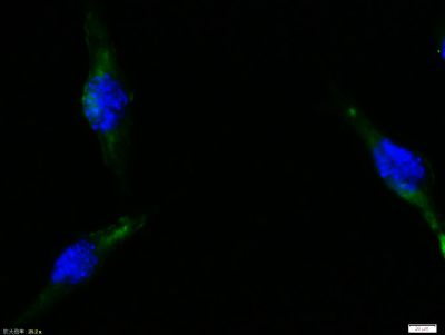
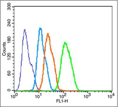
| 产品图片 |  Sample: Sample:Cytochrome C protein at 30 ug Primary: Anti- Cytochrome C (bs-0013R) at 1/300 dilution Secondary: IRDye800CW Goat Anti-Rabbit IgG at 1/20000 dilution Predicted band size: 14 kD Observed band size: 14 kD  Tissue/cell:Sh-sy5y cell; 4% Paraformaldehyde-fixed; Triton X-100 at room temperature for 20 min; Blocking buffer (normal goat serum, C-0005) at 37°C for 20 min; Antibody incubation with (Cytochrome C) polyclonal Antibody, Unconjugated (bs-0013R) 1:100, 90 minutes at 37°C; followed by a FITC conjugated Goat Anti-Rabbit IgG antibody at 37°C for 90 minutes, DAPI (blue, C02-04002) was used to stain the cell nuclei. Tissue/cell:Sh-sy5y cell; 4% Paraformaldehyde-fixed; Triton X-100 at room temperature for 20 min; Blocking buffer (normal goat serum, C-0005) at 37°C for 20 min; Antibody incubation with (Cytochrome C) polyclonal Antibody, Unconjugated (bs-0013R) 1:100, 90 minutes at 37°C; followed by a FITC conjugated Goat Anti-Rabbit IgG antibody at 37°C for 90 minutes, DAPI (blue, C02-04002) was used to stain the cell nuclei. Blank control: HepG2(blue). Blank control: HepG2(blue).Primary Antibody:Rabbit Anti-Cytochrome C antibody (bs-0013R,Green); Dilution: 1μg in 100 μL 1X PBS containing 0.5% BSA; Isotype Control Antibody: Rabbit IgG(orange) ,used under the same conditions; Secondary Antibody: Goat anti-rabbit IgG-FITC(white blue), Dilution: 1:200 in 1 X PBS containing 0.5% BSA. Protocol The cells were fixed with 2% paraformaldehyde for 10 min at 37℃. Primary antibody (bs-0013R, 1μg /1x10^6 cells) were incubated for 30 min at room temperature, followed by 1 X PBS containing 0.5% BSA + 1 0% goat serum (15 min) to block non-specific protein-protein interactions. Then the Goat Anti-rabbit IgG/FITC antibody was added into the blocking buffer mentioned above to react with the primary antibody at 1/200 dilution for 40 min at room temperature. Acquisition of 20,000 events was performed. |