
上海细胞库
人源细胞系| 稳转细胞系| 基因敲除株| 基因点突变细胞株| 基因过表达细胞株| 重组细胞系| 猪的细胞系| 马细胞系| 兔的细胞系| 犬的细胞系| 山羊的细胞系| 鱼的细胞系| 猴的细胞系| 仓鼠的细胞系| 狗的细胞系| 牛的细胞| 大鼠细胞系| 小鼠细胞系| 其他细胞系|
人源细胞系| 稳转细胞系| 基因敲除株| 基因点突变细胞株| 基因过表达细胞株| 重组细胞系| 猪的细胞系| 马细胞系| 兔的细胞系| 犬的细胞系| 山羊的细胞系| 鱼的细胞系| 猴的细胞系| 仓鼠的细胞系| 狗的细胞系| 牛的细胞| 大鼠细胞系| 小鼠细胞系| 其他细胞系|

| 规格 | 价格 | 库存 |
|---|---|---|
| 100ul | ¥ 1680 | 200 |
| 200ul | ¥ 2480 | 200 |
| 中文名称 | Ki67蛋白抗体 |
| 别 名 | Antigen identified by monoclonal antibody Ki 67; Antigen KI67; KIA; Ki-67; Ki67; MKI67; Proliferation related Ki 67 antigen; Antigen KI-67; KI67_HUMAN. |
| 研究领域 | 肿瘤 细胞生物 信号转导 转录调节因子 |
| 抗体来源 | Rabbit |
| 克隆类型 | Polyclonal |
| 交叉反应 | Human, Mouse, |
| 产品应用 | WB=1:500-2000 ELISA=1:500-1000 Flow-Cyt=1ug/test ICC=1:100 not yet tested in other applications. optimal dilutions/concentrations should be determined by the end user. |
| 分 子 量 | 358kDa |
| 细胞定位 | 细胞核 |
| 性 状 | Liquid |
| 浓 度 | 1mg/ml |
| 免 疫 原 | KLH conjugated synthetic peptide derived from human Ki-67:2901-3000/3256 |
| 亚 型 | IgG |
| 纯化方法 | affinity purified by Protein A |
| 储 存 液 | 0.01M TBS(pH7.4) with 1% BSA, 0.03% Proclin300 and 50% Glycerol. |
| 保存条件 | Shipped at 4℃. Store at -20 °C for one year. Avoid repeated freeze/thaw cycles. |
| PubMed | PubMed |
| 产品介绍 | Ki67 antigen is the prototypic cell cycle related nuclear protein, expressed by proliferating cells in all phases of the active cell cycle (G1, S, G2 and M phase). It is absent in resting (G0) cells. Ki67 antibodies are useful in establishing the cell growing fraction in neoplasms (immunohistochemically quantified by determining the number of Ki67 positive cells among the total number of resting cells = Ki67 index). In neoplastic tissues the prognostic value is comparable to the tritiated thymidine labelling index. The correlation between low Ki67 index and histologically low grade tumours is strong. Ki67 is routinely used as a neuronal marker of cell cycling and proliferation. Function: Thought to be required for maintaining cell proliferation. Subcellular Location: Nucleus. Chromosome. Predominantly localized in the G1 phase in the perinucleolar region, in the later phases it is also detected throughout the nuclear interior, being predominantly localized in the nuclear matrix. In mitosis, it is present on all chromosomes. Similarity: Contains 1 FHA domain. SWISS: P46013 Gene ID: 4288 Database links: Entrez Gene: 4288 Human Entrez Gene: 17345 Mouse Entrez Gene: 246042 Rat Omim: 176741 Human SwissProt: P46013 Human SwissProt: Q91VE6 Mouse SwissProt: Q5RJM0 Rat Unigene: 689823 Human Unigene: 80976 Human Unigene: 4078 Mouse Unigene: 233802 Rat Important Note: This product as supplied is intended for research use only, not for use in human, therapeutic or diagnostic applications. 细胞增殖标志物(Proliferation Marker) Ki67与PCNA一样,为细胞增殖的一种标记,在细胞凋亡中S、G2 、M期均有表达,G0期缺如。 Ki-67增殖指数高低与许多肿瘤的分化程度、浸润、转移以及预后密切相关,因此被广泛作为各种恶性肿瘤的必检项目之一。 |
| 产品图片 | 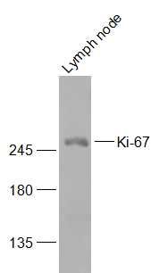 Sample: Sample:Lymph node (Mouse) Lysate at 40 ug Primary: Anti-Ki-67 (bs-23102R) at 1/300 dilution Secondary: IRDye800CW Goat Anti-Rabbit IgG at 1/20000 dilution Predicted band size: 358 kD Observed band size: 358 kD 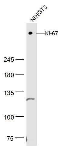 Sample: Sample:NIH/3T3 Cell Lysate at 30 ug Primary: Anti-Ki-67 (bs-23102R) at 1/300 dilution Secondary: IRDye800CW Goat Anti-Rabbit IgG at 1/20000 dilution Predicted band size: 358 kD Observed band size: 358 kD  Tissue/cell: Hela cell; 4% Paraformaldehyde-fixed; Triton X-100 at room temperature for 20 min; Blocking buffer (normal goat serum, C-0005) at 37°C for 20 min; Antibody incubation with (Ki-67) polyclonal Antibody, Unconjugated (bs-23102R) 1:100, 90 minutes at 37°C; followed by a conjugated Goat Anti-Rabbit IgG-AF488 antibody at 37°C for 90 minutes, DAPI (blue, C02-04002) was used to stain the cell nuclei. Tissue/cell: Hela cell; 4% Paraformaldehyde-fixed; Triton X-100 at room temperature for 20 min; Blocking buffer (normal goat serum, C-0005) at 37°C for 20 min; Antibody incubation with (Ki-67) polyclonal Antibody, Unconjugated (bs-23102R) 1:100, 90 minutes at 37°C; followed by a conjugated Goat Anti-Rabbit IgG-AF488 antibody at 37°C for 90 minutes, DAPI (blue, C02-04002) was used to stain the cell nuclei.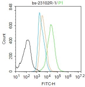 Blank control:HL-60. Blank control:HL-60.Primary Antibody (green line): Rabbit Anti-Ki-67 antibody (bs-23102R) Dilution: 1μg /10^6 cells; Isotype Control Antibody (orange line): Rabbit IgG . Secondary Antibody : Goat anti-rabbit IgG-AF488 Dilution: 1μg /test. Protocol The cells were fixed with 4% PFA (10min at room temperature)and then permeabilized with 90% ice-cold methanol for 20 min at-20℃. The cells were then incubated in 5%BSA to block non-specific protein-protein interactions for 30 min at room temperature .Cells stained with Primary Antibody for 30 min at room temperature. The secondary antibody used for 40 min at room temperature. Acquisition of 20,000 events was performed. 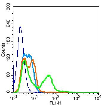 Blank control(blue): HepG2(fixed with 2% paraformaldehyde for 10 min at 37℃). Blank control(blue): HepG2(fixed with 2% paraformaldehyde for 10 min at 37℃).Primary Antibody:Rabbit Anti-Ki-67 antibody (bs-23102R,Green); Dilution: 1μg in 100 μL 1X PBS containing 0.5% BSA; Isotype Control Antibody: Rabbit IgG(orange) ,used under the same conditions; Secondary Antibody: Goat anti-rabbit IgG-FITC(white blue), Dilution: 1:200 in 1 X PBS containing 0.5% BSA. |


