上海细胞库
人源细胞系| 稳转细胞系| 基因敲除株| 基因点突变细胞株| 基因过表达细胞株| 重组细胞系| 猪的细胞系| 马细胞系| 兔的细胞系| 犬的细胞系| 山羊的细胞系| 鱼的细胞系| 猴的细胞系| 仓鼠的细胞系| 狗的细胞系| 牛的细胞| 大鼠细胞系| 小鼠细胞系| 其他细胞系|

| 规格 | 价格 | 库存 |
|---|---|---|
| 100ul | ¥ 1980 | 200 |
| 中文名称 | 磷酸化丝氨酸/苏氨酸蛋白激酶MAK抗体 |
| 别 名 | MAK (phospho Tyr159); MAK (phospho Y159); dJ417M14.2; Mak; MAK_HUMAN; Male germ cell associated kinase; Male germ cell-associated kinase; OTTHUMP00000016025; Rck; Serine/threonine protein kinase MAK; Serine/threonine-protein kinase MAK. |
| 产品类型 | 磷酸化抗体 |
| 研究领域 | 细胞生物 发育生物学 信号转导 激酶和磷酸酶 |
| 抗体来源 | Rabbit |
| 克隆类型 | Polyclonal |
| 交叉反应 | Human, Mouse, Rat, |
| 产品应用 | WB=1:500-2000 ELISA=1:500-1000 IHC-P=1:100-500 IHC-F=1:100-500 ICC=1:100-500 IF=1:100-500 (石蜡切片需做抗原修复) not yet tested in other applications. optimal dilutions/concentrations should be determined by the end user. |
| 分 子 量 | 71kDa |
| 细胞定位 | 细胞核 细胞浆 |
| 性 状 | Liquid |
| 浓 度 | 1mg/ml |
| 免 疫 原 | KLH conjugated synthesised phosphopeptide derived from human MAK around the phosphorylation site of Tyr159:TD(p-Y)VS |
| 亚 型 | IgG |
| 纯化方法 | affinity purified by Protein A |
| 储 存 液 | 0.01M TBS(pH7.4) with 1% BSA, 0.03% Proclin300 and 50% Glycerol. |
| 保存条件 | Shipped at 4℃. Store at -20 °C for one year. Avoid repeated freeze/thaw cycles. |
| PubMed | PubMed |
| 产品介绍 | The product of this gene is a serine/threonine protein kinase related to kinases involved in cell cycle regulation. It is expressed almost exclusively in the testis, primarily in germ cells. Studies of the mouse and rat homologs have localized the kinase to the chromosomes during meiosis in spermatogenesis, specifically to the synaptonemal complex that exists while homologous chromosomes are paired. There is, however, a study of the mouse homolog that has identified high levels of expression in developing sensory epithelia so its function may be more generalized. Three transcript variants encoding different isoforms have been found for this gene. [provided by RefSeq, Jul 2011] Function: Essential for the regulation of ciliary length and required for the long-term survival of photoreceptors (By similarity). Phosphorylates FZR1 in a cell cycle-dependent manner. Plays a role in the transcriptional coactivation of AR. Could play an important function in spermatogenesis. May play a role in chromosomal stability in prostate cancer cells. Subcellular Location: Nucleus. Cytoplasm > cytoskeleton > centrosome. Cytoplasm > cytoskeleton > spindle. Midbody. Cell projection > cilium > photoreceptor outer segment. Photoreceptor inner segment. Localized in both the connecting cilia and the outer segment axonemes (By similarity). Localized uniformly in nuclei during interphase, to the mitotic spindle and centrosomes during metaphase and anaphase, and also to midbody at anaphase until telophase. Tissue Specificity: Expressed in prostate cancer cell lines at generally higher levels than in normal prostate epithelial cell lines. Isoform 1 is expressed in kidney, testis, lung, trachea, and retina. Isoform 2 is retina-specific where it is expressed in rod and cone photoreceptors. Post-translational modifications: Autophosphorylated. Phosphorylated on serine and threonine residues. DISEASE: Defects in MAK are the cause of retinitis pigmentosa type 62 (RP62) [MIM:614181]. RP62 is a retinal dystrophy belonging to the group of pigmentary retinopathies. Retinitis pigmentosa is characterized by retinal pigment deposits visible on fundus examination and primary loss of rod photoreceptor cells followed by secondary loss of cone photoreceptors. Patients typically have night vision blindness and loss of midperipheral visual field. As their condition progresses, they lose their far peripheral visual field and eventually central vision as well. Similarity: Belongs to the protein kinase superfamily. CMGC Ser/Thr protein kinase family. CDC2/CDKX subfamily. Contains 1 protein kinase domain. SWISS: P20794 Gene ID: 4117 Database links: Entrez Gene: 4117 Human Entrez Gene: 17152 Mouse Entrez Gene: 25677 Rat Omim: 154235 Human SwissProt: P20794 Human SwissProt: Q04859 Mouse SwissProt: P20793 Rat Unigene: 446125 Human Unigene: 8149 Mouse Unigene: 9670 Rat Important Note: This product as supplied is intended for research use only, not for use in human, therapeutic or diagnostic applications. |
| 产品图片 | 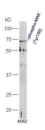 Protein: k562(human) lysate at 40ug; Protein: k562(human) lysate at 40ug;Primary: rabbit Anti-phospho-MAK (Tyr159) (bs-18634R) at 1:300; Secondary: HRP conjugated Goat-Anti-rabbit IgG(bs-0295G-HRP) at 1: 5000; Predicted band size: 71 kD Observed band size: 71 kD 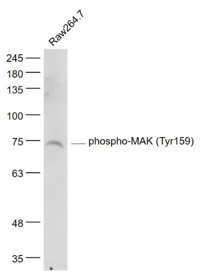 Sample: Sample:Raw264.7(Mouse) Cell Lysate at 30 ug Primary: Anti- phospho-MAK (Tyr159) (bs-18634R) at 1/1000 dilution Secondary: IRDye800CW Goat Anti-Rabbit IgG at 1/20000 dilution Predicted band size: 71 kD Observed band size: 73 kD 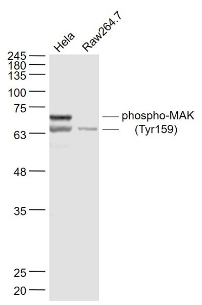 Sample: Sample:Hela(Human) Cell Lysate at 30 ug Raw264.7(Mouse) Cell Lysate at 30 ug Primary: Anti- phospho-MAK (Tyr159) (bs-18634R) at 1/1000 dilution Secondary: IRDye800CW Goat Anti-Rabbit IgG at 1/20000 dilution Predicted band size: 71 kD Observed band size: 71/66 kD 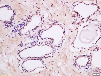 Tissue/cell: human prostate tissue; 4% Paraformaldehyde-fixed and paraffin-embedded; Tissue/cell: human prostate tissue; 4% Paraformaldehyde-fixed and paraffin-embedded;Antigen retrieval: citrate buffer ( 0.01M, pH 6.0 ), Boiling bathing for 15min; Block endogenous peroxidase by 3% Hydrogen peroxide for 30min; Blocking buffer (normal goat serum,C-0005) at 37℃ for 20 min; Incubation: Anti-phospho-MAK (Tyr159) Polyclonal Antibody, Unconjugated(bs-18634R) 1:200, overnight at 4°C, followed by conjugation to the secondary antibody(SP-0023) and DAB(C-0010) staining 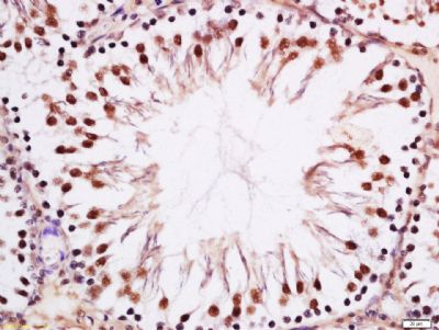 Tissue/cell: rat testis tissue; 4% Paraformaldehyde-fixed and paraffin-embedded; Tissue/cell: rat testis tissue; 4% Paraformaldehyde-fixed and paraffin-embedded;Antigen retrieval: citrate buffer ( 0.01M, pH 6.0 ), Boiling bathing for 15min; Block endogenous peroxidase by 3% Hydrogen peroxide for 30min; Blocking buffer (normal goat serum,C-0005) at 37℃ for 20 min; Incubation: Anti-phospho-MAK (Tyr159) Polyclonal Antibody, Unconjugated(bs-18634R) 1:200, overnight at 4°C, followed by conjugation to the secondary antibody(SP-0023) and DAB(C-0010) staining |