上海细胞库
人源细胞系| 稳转细胞系| 基因敲除株| 基因点突变细胞株| 基因过表达细胞株| 重组细胞系| 猪的细胞系| 马细胞系| 兔的细胞系| 犬的细胞系| 山羊的细胞系| 鱼的细胞系| 猴的细胞系| 仓鼠的细胞系| 狗的细胞系| 牛的细胞| 大鼠细胞系| 小鼠细胞系| 其他细胞系|

| 规格 | 价格 | 库存 |
|---|---|---|
| 100ul | ¥ 1680 | 200 |
| 200ul | ¥ 2480 | 200 |
| 中文名称 | 单丝氨酸蛋白激酶1+2抗体 |
| 别 名 | LIM kinase 1+2; LIMK1+LIMK2; LIM domain kinase 1; LIM domain kinase 2; LIMK1; LIMK2;LIMK1_HUMAN; LIMK2_HUMAN. |
| 研究领域 | 细胞生物 神经生物学 信号转导 细胞骨架 表观遗传学 |
| 抗体来源 | Rabbit |
| 克隆类型 | Polyclonal |
| 交叉反应 | Mouse, Rat, (predicted: Human, Dog, Pig, Horse, Rabbit, Sheep, ) |
| 产品应用 | WB=1:500-2000 ELISA=1:500-1000 IHC-P=1:100-500 IHC-F=1:100-500 ICC=1:100-500 IF=1:200-800 (石蜡切片需做抗原修复) not yet tested in other applications. optimal dilutions/concentrations should be determined by the end user. |
| 分 子 量 | 72, 73kDa |
| 细胞定位 | 细胞核 细胞浆 |
| 性 状 | Liquid |
| 浓 度 | 1mg/ml |
| 免 疫 原 | KLH conjugated synthetic peptide derived from human LIM kinase 1 + 2:365-470/647 |
| 亚 型 | IgG |
| 纯化方法 | affinity purified by Protein A |
| 储 存 液 | 0.01M TBS(pH7.4) with 1% BSA, 0.03% Proclin300 and 50% Glycerol. |
| 保存条件 | Shipped at 4℃. Store at -20 °C for one year. Avoid repeated freeze/thaw cycles. |
| PubMed | PubMed |
| 产品介绍 | LIMK 1 and 2 likely regulate aspects of the cytoskeleton, through control of the organization of actin filaments. They can phosphorylate an actin-binding protein, cofilin which binds to actin monomers and polymers and promotes the disassembly of actin filament.The phosphorylation of cofilin via LIMK inactivates this potential. LIMK1 is highly active in the brain and spinal chord, where it is believed to be involved in the development of nerve cells whilst LIMK2 is ubiquitously expressed in many adult tissues. LIMK1 may play an important role in areas of the brain that are responsible for processing visual-spatial information (visuospatial constructive cognition). These parts of the brain are important for visualizing an object as a set of parts and performing tasks such as writing, drawing, constructing models, and assembling puzzles. LIMK1 is specifically stimulated by Rac, one of the Rho family proteins, while LIMK2 activity is activated under the control of other Rho family members, Rho and Cdc42, suggesting that two distinct pathways exist in the Rho family driven actin cytoskeleton dynamics. Function: Protein kinase which regulates actin filament dynamics. Phosphorylates and inactivates the actin binding/depolymerizing factor cofilin, thereby stabilizing the actin cytoskeleton. Stimulates axonal outgrowth and may be involved in brain development. Isoform 3 has a dominant negative effect on actin cytoskeletal changes. Subunit: Interacts (via LIM domain) with the cytoplasmic domain of NRG1 (By similarity). Interacts with NISCH (By similarity). Interacts with RLIM and RNF6 (By similarity). Self-associates to form homodimers. Interacts with HSP90AA1; this interaction promotes LIMK1 dimerization and subsequent transphosphorylation. Interacts with CDN1C. Interacts with SSH1. Interacts with ROCK1. Subcellular Location: LIMK1 Cytoplasmic LIMK2 Cytoplasmic and Nuclear. Isoform LIMK2a is distributed in the cytoplasm and the nucleus, and isoform LIMK2b occurs mainly in the cytoplasm and is scarcely translocated to the nucleus. Tissue Specificity: Highest expression in both adult and fetal nervous system. Detected ubiquitously throughout the different regions of adult brain, with highest levels in the cerebral cortex. Expressed to a lesser extent in heart and skeletal muscle. Post-translational modifications: Autophosphorylated (By similarity). Phosphorylated on Thr-508 by ROCK1 and PAK1, resulting in activation. Phosphorylated by PAK4 which increases the ability of LIMK1 to phosphorylate cofilin. Phosphorylated at Ser-323 by MAPKAPK2 during activation of VEGFA-induced signaling, which results in activation of LIMK1 and promotion of actin reorganization, cell migration, and tubule formation of endothelial cells. Dephosphorylated and inactivated by SSH1. Phosphorylated by CDC42BP. Ubiquitinated. 'Lys-48'-linked polyubiquitination by RNF6 leads to proteasomal degradation through the 26S proteasome, modulating LIMK1 levels in the growth cone and its effect on axonal outgrowth. Also polyubiquitinated by RLIM DISEASE: Note=LIMK1 is located in the Williams-Beuren syndrome (WBS) critical region. WBS results from a hemizygous deletion of several genes on chromosome 7q11.23, thought to arise as a consequence of unequal crossing over between highly homologous low-copy repeat sequences flanking the deleted region. Similarity: Belongs to the protein kinase superfamily. TKL Ser/Thr protein kinase family. Contains 2 LIM zinc-binding domains. Contains 1 PDZ (DHR) domain. Contains 1 protein kinase domain. SWISS: P53667 Gene ID: 3984 Database links: Entrez Gene: 3984 Human Entrez Gene: 16885 Mouse Entrez Gene: 65172 Rat Omim: 601329 Human SwissProt: P53667 Human SwissProt: P53668 Mouse SwissProt: P53669 Rat Unigene: 647035 Human Unigene: 15409 Mouse Unigene: 11250 Rat Important Note: This product as supplied is intended for research use only, not for use in human, therapeutic or diagnostic applications. |
| 产品图片 | 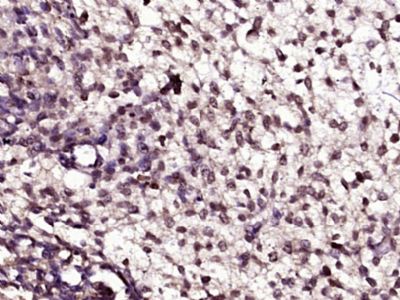 Paraformaldehyde-fixed, paraffin embedded (Mouse embryos); Antigen retrieval by boiling in sodium citrate buffer (pH6.0) for 15min; Block endogenous peroxidase by 3% hydrogen peroxide for 20 minutes; Blocking buffer (normal goat serum) at 37°C for 30min; Antibody incubation with (LIM kinase 1 + 2) Polyclonal Antibody, Unconjugated (bs-11871R) at 1:400 overnight at 4°C, followed by operating according to SP Kit(Rabbit) (sp-0023) instructionsand DAB staining. Paraformaldehyde-fixed, paraffin embedded (Mouse embryos); Antigen retrieval by boiling in sodium citrate buffer (pH6.0) for 15min; Block endogenous peroxidase by 3% hydrogen peroxide for 20 minutes; Blocking buffer (normal goat serum) at 37°C for 30min; Antibody incubation with (LIM kinase 1 + 2) Polyclonal Antibody, Unconjugated (bs-11871R) at 1:400 overnight at 4°C, followed by operating according to SP Kit(Rabbit) (sp-0023) instructionsand DAB staining.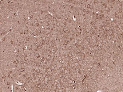 Paraformaldehyde-fixed, paraffin embedded (Mouse brain); Antigen retrieval by boiling in sodium citrate buffer (pH6.0) for 15min; Block endogenous peroxidase by 3% hydrogen peroxide for 20 minutes; Blocking buffer (normal goat serum) at 37°C for 30min; Antibody incubation with (LIM kinase 1 + 2) Polyclonal Antibody, Unconjugated (bs-11871R) at 1:400 overnight at 4°C, followed by operating according to SP Kit(Rabbit) (sp-0023) instructionsand DAB staining. Paraformaldehyde-fixed, paraffin embedded (Mouse brain); Antigen retrieval by boiling in sodium citrate buffer (pH6.0) for 15min; Block endogenous peroxidase by 3% hydrogen peroxide for 20 minutes; Blocking buffer (normal goat serum) at 37°C for 30min; Antibody incubation with (LIM kinase 1 + 2) Polyclonal Antibody, Unconjugated (bs-11871R) at 1:400 overnight at 4°C, followed by operating according to SP Kit(Rabbit) (sp-0023) instructionsand DAB staining.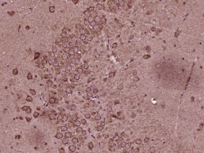 Paraformaldehyde-fixed, paraffin embedded (Rat brain); Antigen retrieval by boiling in sodium citrate buffer (pH6.0) for 15min; Block endogenous peroxidase by 3% hydrogen peroxide for 20 minutes; Blocking buffer (normal goat serum) at 37°C for 30min; Antibody incubation with (LIM kinase 1 + 2) Polyclonal Antibody, Unconjugated (bs-11871R) at 1:400 overnight at 4°C, followed by operating according to SP Kit(Rabbit) (sp-0023) instructionsand DAB staining. Paraformaldehyde-fixed, paraffin embedded (Rat brain); Antigen retrieval by boiling in sodium citrate buffer (pH6.0) for 15min; Block endogenous peroxidase by 3% hydrogen peroxide for 20 minutes; Blocking buffer (normal goat serum) at 37°C for 30min; Antibody incubation with (LIM kinase 1 + 2) Polyclonal Antibody, Unconjugated (bs-11871R) at 1:400 overnight at 4°C, followed by operating according to SP Kit(Rabbit) (sp-0023) instructionsand DAB staining.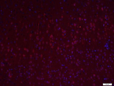 Paraformaldehyde-fixed, paraffin embedded (Mouse brain); Antigen retrieval by boiling in sodium citrate buffer (pH6.0) for 15min; Block endogenous peroxidase by 3% hydrogen peroxide for 20 minutes; Blocking buffer (normal goat serum) at 37°C for 30min; Antibody incubation with (LIM kinase 1 + 2) Polyclonal Antibody, Unconjugated (bs-11871R) at 1:400 overnight at 4°C, followed by a conjugated Goat Anti-Rabbit IgG antibody (bs-0295G-CY3) for 90 minutes, and DAPI for nuclei staining. Paraformaldehyde-fixed, paraffin embedded (Mouse brain); Antigen retrieval by boiling in sodium citrate buffer (pH6.0) for 15min; Block endogenous peroxidase by 3% hydrogen peroxide for 20 minutes; Blocking buffer (normal goat serum) at 37°C for 30min; Antibody incubation with (LIM kinase 1 + 2) Polyclonal Antibody, Unconjugated (bs-11871R) at 1:400 overnight at 4°C, followed by a conjugated Goat Anti-Rabbit IgG antibody (bs-0295G-CY3) for 90 minutes, and DAPI for nuclei staining.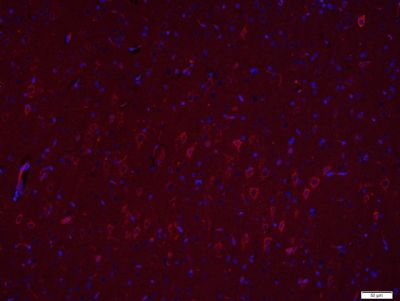 Paraformaldehyde-fixed, paraffin embedded (Rat brain); Antigen retrieval by boiling in sodium citrate buffer (pH6.0) for 15min; Block endogenous peroxidase by 3% hydrogen peroxide for 20 minutes; Blocking buffer (normal goat serum) at 37°C for 30min; Antibody incubation with (LIM kinase 1 + 2) Polyclonal Antibody, Unconjugated (bs-11871R) at 1:400 overnight at 4°C, followed by a conjugated Goat Anti-Rabbit IgG antibody (bs-0295G-CY3) for 90 minutes, and DAPI for nuclei staining. Paraformaldehyde-fixed, paraffin embedded (Rat brain); Antigen retrieval by boiling in sodium citrate buffer (pH6.0) for 15min; Block endogenous peroxidase by 3% hydrogen peroxide for 20 minutes; Blocking buffer (normal goat serum) at 37°C for 30min; Antibody incubation with (LIM kinase 1 + 2) Polyclonal Antibody, Unconjugated (bs-11871R) at 1:400 overnight at 4°C, followed by a conjugated Goat Anti-Rabbit IgG antibody (bs-0295G-CY3) for 90 minutes, and DAPI for nuclei staining. |