上海细胞库
人源细胞系| 稳转细胞系| 基因敲除株| 基因点突变细胞株| 基因过表达细胞株| 重组细胞系| 猪的细胞系| 马细胞系| 兔的细胞系| 犬的细胞系| 山羊的细胞系| 鱼的细胞系| 猴的细胞系| 仓鼠的细胞系| 狗的细胞系| 牛的细胞| 大鼠细胞系| 小鼠细胞系| 其他细胞系|

| 规格 | 价格 | 库存 |
|---|---|---|
| 100ul | ¥ 1680 | 200 |
| 200ul | ¥ 2480 | 200 |
| 中文名称 | 嘌呤嘧啶核酸内切酶2抗体 |
| 别 名 | AP endonuclease 2; AP endonuclease XTH2; APE 2; APE2; APEX 2; APEX L2; APEX nuclease (apurinic/apyrimidinic endonuclease) 2; APEX Nuclease 2; APEX nuclease like 2; APEXL 2; APEXL2; Apurinic apyrimidinic endonuclease 2; Apurinic/apyrimidinic endonuclease 2; Apurinic/apyrimidinic endonuclease like 2; C430040P13Rik; DNA (apurinic or apyrimidinic site) lyase 2; XTH 2; XTH2; APEX2_HUMAN. |
| 研究领域 | 细胞生物 染色质和核信号 |
| 抗体来源 | Rabbit |
| 克隆类型 | Polyclonal |
| 交叉反应 | Mouse, Rat, (predicted: Human, Dog, Cow, ) |
| 产品应用 | WB=1:500-2000 ELISA=1:500-1000 IHC-P=1:100-500 IHC-F=1:100-500 IF=1:100-500 (石蜡切片需做抗原修复) not yet tested in other applications. optimal dilutions/concentrations should be determined by the end user. |
| 分 子 量 | 57kDa |
| 细胞定位 | 细胞核 细胞浆 |
| 性 状 | Liquid |
| 浓 度 | 1mg/ml |
| 免 疫 原 | KLH conjugated synthetic peptide derived from human APEX2:301-200/518 |
| 亚 型 | IgG |
| 纯化方法 | affinity purified by Protein A |
| 储 存 液 | 0.01M TBS(pH7.4) with 1% BSA, 0.03% Proclin300 and 50% Glycerol. |
| 保存条件 | Shipped at 4℃. Store at -20 °C for one year. Avoid repeated freeze/thaw cycles. |
| PubMed | PubMed |
| 产品介绍 | Apurinic/apyrimidinic (AP) sites occur frequently in DNA molecules by spontaneous hydrolysis, by DNA damaging agents or by DNA glycosylases that remove specific abnormal bases. AP sites are pre-mutagenic lesions that can prevent normal DNA replication so the cell contains systems to identify and repair such sites. Class II AP endonucleases cleave the phosphodiester backbone 5' to the AP site. This gene encodes a protein shown to have a weak class II AP endonuclease activity. Most of the encoded protein is located in the nucleus but some is also present in mitochondria. This protein may play an important role in both nuclear and mitochondrial base excision repair (BER). Function: Function as a weak apurinic/apyrimidinic (AP) endodeoxyribonuclease in the DNA base excision repair (BER) pathway of DNA lesions induced by oxidative and alkylating agents. Initiates repair of AP sites in DNA by catalyzing hydrolytic incision of the phosphodiester backbone immediately adjacent to the damage, generating a single-strand break with 5'-deoxyribose phosphate and 3'-hydroxyl ends. Displays also double-stranded DNA 3'-5' exonuclease, 3'-phosphodiesterase activities. Shows robust 3'-5' exonuclease activity on 3'-recessed heteroduplex DNA and is able to remove mismatched nucleotides preferentially. Shows fairly strong 3'-phosphodiesterase activity involved in the removal of 3'-damaged termini formed in DNA by oxidative agents. In the nucleus functions in the PCNA-dependent BER pathway. Required for somatic hypermutation (SHM) and DNA cleavage step of class switch recombination (CSR) of immunoglobulin genes. Required for proper cell cycle progression during proliferation of peripheral lymphocytes. Subunit: Interacts with PCNA; this interaction is triggered by reactive oxygen species and increased by misincorporation of uracil in nuclear DNA. Subcellular Location: Nucleus. Cytoplasm. Mitochondrion (Probable). Note=Together with PCNA, is redistributed in discrete nuclear foci in presence of oxidative DNA damaging agents. Tissue Specificity: Highly expressed in brain and kidney. Weakly expressed in the fetal brain. Similarity: Belongs to the DNA repair enzymes AP/ExoA family. SWISS: Q9UBZ4 Gene ID: 27301 Database links: Entrez Gene: Human Entrez Gene: 77622 Mouse Entrez Gene: 289662 Rat Omim: 300773 Human SwissProt: Q9UBZ4 Human SwissProt: Q68G58 Mouse Unigene: 659558 Human Unigene: 440275 Mouse Important Note: This product as supplied is intended for research use only, not for use in human, therapeutic or diagnostic applications. |
| 产品图片 | 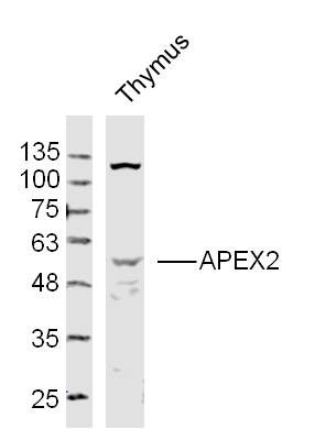 Sample: Sample:Thymus (Mouse) Lysate at 40 ug Primary: Anti- APEX2 (bs-6587R)at 1/300 dilution Secondary: IRDye800CW Goat Anti-Rabbit IgG at 1/20000 dilution Predicted band size: 57kD Observed band size: 57kD 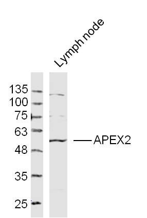 Sample: Sample:Lymph node(Mouse) Lysate at 40 ug Primary: Anti- APEX2 (bs-6587R)at 1/300 dilution Secondary: IRDye800CW Goat Anti-Rabbit IgG at 1/20000 dilution Predicted band size: 57kD Observed band size: 57kD 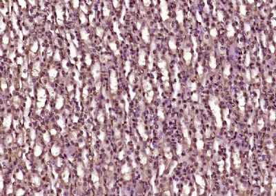 Paraformaldehyde-fixed, paraffin embedded (Mouse kidney); Antigen retrieval by boiling in sodium citrate buffer (pH6.0) for 15min; Block endogenous peroxidase by 3% hydrogen peroxide for 20 minutes; Blocking buffer (normal goat serum) at 37°C for 30min; Antibody incubation with (APEX2) Polyclonal Antibody, Unconjugated (bs-6587R) at 1:400 overnight at 4°C, followed by operating according to SP Kit(Rabbit) (sp-0023) instructionsand DAB staining. Paraformaldehyde-fixed, paraffin embedded (Mouse kidney); Antigen retrieval by boiling in sodium citrate buffer (pH6.0) for 15min; Block endogenous peroxidase by 3% hydrogen peroxide for 20 minutes; Blocking buffer (normal goat serum) at 37°C for 30min; Antibody incubation with (APEX2) Polyclonal Antibody, Unconjugated (bs-6587R) at 1:400 overnight at 4°C, followed by operating according to SP Kit(Rabbit) (sp-0023) instructionsand DAB staining.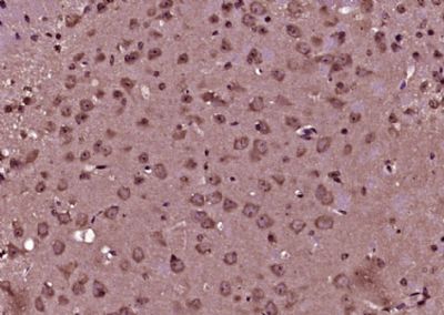 Paraformaldehyde-fixed, paraffin embedded (Mouse brain); Antigen retrieval by boiling in sodium citrate buffer (pH6.0) for 15min; Block endogenous peroxidase by 3% hydrogen peroxide for 20 minutes; Blocking buffer (normal goat serum) at 37°C for 30min; Antibody incubation with (APEX2) Polyclonal Antibody, Unconjugated (bs-6587R) at 1:400 overnight at 4°C, followed by operating according to SP Kit(Rabbit) (sp-0023) instructionsand DAB staining. Paraformaldehyde-fixed, paraffin embedded (Mouse brain); Antigen retrieval by boiling in sodium citrate buffer (pH6.0) for 15min; Block endogenous peroxidase by 3% hydrogen peroxide for 20 minutes; Blocking buffer (normal goat serum) at 37°C for 30min; Antibody incubation with (APEX2) Polyclonal Antibody, Unconjugated (bs-6587R) at 1:400 overnight at 4°C, followed by operating according to SP Kit(Rabbit) (sp-0023) instructionsand DAB staining.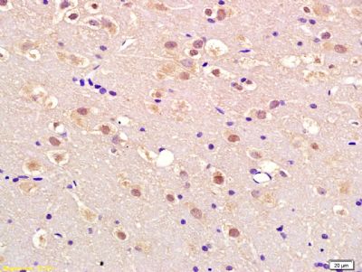 Tissue/cell: rat brain tissue; 4% Paraformaldehyde-fixed and paraffin-embedded; Tissue/cell: rat brain tissue; 4% Paraformaldehyde-fixed and paraffin-embedded;Antigen retrieval: citrate buffer ( 0.01M, pH 6.0 ), Boiling bathing for 15min; Block endogenous peroxidase by 3% Hydrogen peroxide for 30min; Blocking buffer (normal goat serum,C-0005) at 37℃ for 20 min; Incubation: Anti-APEX2 Polyclonal Antibody, Unconjugated(bs-6587R) 1:200, overnight at 4°C, followed by conjugation to the secondary antibody(SP-0023) and DAB(C-0010) staining |