上海细胞库
人源细胞系| 稳转细胞系| 基因敲除株| 基因点突变细胞株| 基因过表达细胞株| 重组细胞系| 猪的细胞系| 马细胞系| 兔的细胞系| 犬的细胞系| 山羊的细胞系| 鱼的细胞系| 猴的细胞系| 仓鼠的细胞系| 狗的细胞系| 牛的细胞| 大鼠细胞系| 小鼠细胞系| 其他细胞系|

| 规格 | 价格 | 库存 |
|---|---|---|
| 100ul | ¥ 1980 | 200 |
| 中文名称 | 磷酸化丝裂原活化蛋白激酶激酶8抗体 |
| 别 名 | MAP3K8 (phospho S400); P-MAP3K8/Tpl2 (Ser400); Phospho-MAP3K8(Ser400); Phospho-Tpl2 (Ser400); P-Tpl2 (Ser400);c COT; Cancer Osaka thyroid oncogene; Cancer Osaka thyroid oncogene; CCOT; COT; COT proto oncogene serine/threonine protein kinase; EST; ESTF; Ewing sarcoma transformant; FLJ10486; M3K8_HUMAN; MAP3K 8; MAP3K8; Mitogen activated protein kinase kinase kinase 8; Mitogen-activated protein kinase kinase kinase 8; Proto oncogene cCot; Proto-oncogene c-Cot; Serine/threonine protein kinase cot; Serine/threonine-protein kinase cot; TPL 2; TPL-2; TPL2; Tumor progression locus 2. |
| 产品类型 | 磷酸化抗体 |
| 研究领域 | 肿瘤 免疫学 信号转导 激酶和磷酸酶 |
| 抗体来源 | Rabbit |
| 克隆类型 | Polyclonal |
| 交叉反应 | Human, Mouse, Rat, (predicted: Chicken, Dog, Pig, Cow, Horse, Rabbit, ) |
| 产品应用 | WB=1:500-2000 ELISA=1:500-1000 IHC-P=1:100-500 IHC-F=1:100-500 Flow-Cyt=1ug/Test IF=1:100-500 (石蜡切片需做抗原修复) not yet tested in other applications. optimal dilutions/concentrations should be determined by the end user. |
| 分 子 量 | 53kDa |
| 细胞定位 | 细胞浆 |
| 性 状 | Liquid |
| 浓 度 | 1mg/ml |
| 免 疫 原 | KLH conjugated Synthesised phosphopeptide derived from human MAP3K8/Tpl2 around the phosphorylation site of Ser400:CQ(p-S)LD |
| 亚 型 | IgG |
| 纯化方法 | affinity purified by Protein A |
| 储 存 液 | 0.01M TBS(pH7.4) with 1% BSA, 0.03% Proclin300 and 50% Glycerol. |
| 保存条件 | Shipped at 4℃. Store at -20 °C for one year. Avoid repeated freeze/thaw cycles. |
| PubMed | PubMed |
| 产品介绍 | This gene is an oncogene that encodes a member of the serine/threonine protein kinase family. The encoded protein localizes to the cytoplasm and can activate both the MAP kinase and JNK kinase pathways. This protein was shown to activate IkappaB kinases, and thus induce the nuclear production of NF-kappaB. This protein was also found to promote the production of TNF-alpha and IL-2 during T lymphocyte activation. This gene may also utilize a downstream in-frame translation start codon, and thus produce an isoform containing a shorter N-terminus. The shorter isoform has been shown to display weaker transforming activity. Alternate splicing results in multiple transcript variants that encode the same protein. [provided by RefSeq, Sep 2011] Function: Required for TLR4 activation of the MEK/ERK pathway. Able to activate NF-kappa-B 1 by stimulating proteasome-mediated proteolysis of NF-kappa-B 1/p105. Plays a role in the cell cycle. The longer form has some transforming activity, although it is much weaker than the activated cot oncoprotein. Subunit: Forms a ternary complex with NFKB1 and TNIP2. Subcellular Location: Cytoplasm. Tissue Specificity: Expressed in several normal tissues and human tumor-derived cell lines. Post-translational modifications: Autophosphorylated. Isoform 1 undergoes phosphorylation mainly on Ser residues, and isoform 2 on both Ser and Thr residues. Similarity: Belongs to the protein kinase superfamily. STE Ser/Thr protein kinase family. MAP kinase kinase kinase subfamily. Contains 1 protein kinase domain. SWISS: P41279 Gene ID: 1326 Database links: Entrez Gene: 1326 Human Entrez Gene: 26410 Mouse Entrez Gene: 116596 Rat Omim: 191195 Human SwissProt: P41279 Human SwissProt: Q07174 Mouse SwissProt: Q63562 Rat Unigene: 432453 Human Unigene: 3275 Mouse Unigene: 9939 Rat Important Note: This product as supplied is intended for research use only, not for use in human, therapeutic or diagnostic applications. |
| 产品图片 | 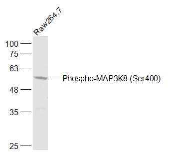 Sample: Sample:RAW264.7(Mouse) Cell Lysate at 30 ug Primary: Anti-Phospho-MAP3K8 (Ser400) (bs-3454R) at 1/1000 dilution Secondary: IRDye800CW Goat Anti-Rabbit IgG at 1/20000 dilution Predicted band size: 53 kD Observed band size: 53 kD 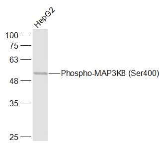 Sample: Sample:HepG2(Human) Cell Lysate at 30 ug Primary: Anti-Phospho-MAP3K8 (Ser400) (bs-3454R) at 1/1000 dilution Secondary: IRDye800CW Goat Anti-Rabbit IgG at 1/20000 dilution Predicted band size: 53 kD Observed band size: 53 kD 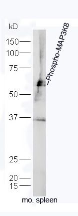 Sample: Sample:Spleen (Mouse) Lysate at 40 ug Primary: Anti-Phospho-MAP3K8 (Ser400) (bs-3454R) at 1/300 dilution Secondary: IRDye800CW Goat Anti-Rabbit IgG at 1/20000 dilution Predicted band size: 53 kD Observed band size: 55 kD 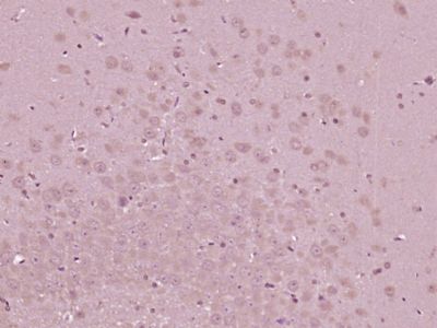 Paraformaldehyde-fixed, paraffin embedded (Rat brain); Antigen retrieval by boiling in sodium citrate buffer (pH6.0) for 15min; Block endogenous peroxidase by 3% hydrogen peroxide for 20 minutes; Blocking buffer (normal goat serum) at 37°C for 30min; Antibody incubation with (Phospho-MAP3K8 (Ser400)) Polyclonal Antibody, Unconjugated (bs-3454R) at 1:400 overnight at 4°C, followed by operating according to SP Kit(Rabbit) (sp-0023) instructionsand DAB staining. Paraformaldehyde-fixed, paraffin embedded (Rat brain); Antigen retrieval by boiling in sodium citrate buffer (pH6.0) for 15min; Block endogenous peroxidase by 3% hydrogen peroxide for 20 minutes; Blocking buffer (normal goat serum) at 37°C for 30min; Antibody incubation with (Phospho-MAP3K8 (Ser400)) Polyclonal Antibody, Unconjugated (bs-3454R) at 1:400 overnight at 4°C, followed by operating according to SP Kit(Rabbit) (sp-0023) instructionsand DAB staining.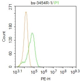 Blank control: Hela. Blank control: Hela.Primary Antibody (green line): Rabbit Anti-MAP3K8 antibody (bs-3454R) Dilution: 1μg /10^6 cells; Isotype Control Antibody (orange line): Rabbit IgG . Secondary Antibody : Goat anti-rabbit IgG-PE Dilution: 1μg /test. Protocol The cells were fixed with 4% PFA (10min at room temperature)and then permeabilized with PBST for 20 min at room temperature. The cells were then incubated in 5%BSA to block non-specific protein-protein interactions for 30 min at at room temperature .Cells stained with Primary Antibody for 30 min at room temperature. The secondary antibody used for 40 min at room temperature. Acquisition of 20,000 events was performed. |