
上海细胞库
人源细胞系| 稳转细胞系| 基因敲除株| 基因点突变细胞株| 基因过表达细胞株| 重组细胞系| 猪的细胞系| 马细胞系| 兔的细胞系| 犬的细胞系| 山羊的细胞系| 鱼的细胞系| 猴的细胞系| 仓鼠的细胞系| 狗的细胞系| 牛的细胞| 大鼠细胞系| 小鼠细胞系| 其他细胞系|
人源细胞系| 稳转细胞系| 基因敲除株| 基因点突变细胞株| 基因过表达细胞株| 重组细胞系| 猪的细胞系| 马细胞系| 兔的细胞系| 犬的细胞系| 山羊的细胞系| 鱼的细胞系| 猴的细胞系| 仓鼠的细胞系| 狗的细胞系| 牛的细胞| 大鼠细胞系| 小鼠细胞系| 其他细胞系|

| 规格 | 价格 | 库存 |
|---|---|---|
| 100ul | ¥ 1880 | 200 |
| 中文名称 | 磷酸化神经生长相关蛋白43抗体 |
| 别 名 | GAP43 (phospho S41); p-GAP43 (phospho S41); Phospho-GAP43(pSer41); GAP43 (Phospho-Ser41); p-GAP43 (Ser41); p-GAP43 (S41); Growth Associated Protein-43; Neuromodulin; Axonal membrane protein GAP 43; B-50; F1; GAP 43; Growth Associated Protein 43; Nerve Growth Related Peptide; Neural phosphoprotein B 50; Neuromodulin; GAP-43; pp46; NEUM_HUMAN; Protein F1; QtrA-11580; QtrA-13071. |
| 产品类型 | 磷酸化抗体 |
| 研究领域 | 细胞生物 免疫学 神经生物学 信号转导 细胞凋亡 转录调节因子 |
| 抗体来源 | Rabbit |
| 克隆类型 | Polyclonal |
| 交叉反应 | Human, Mouse, Rat, (predicted: Dog, ) |
| 产品应用 | ELISA=1:500-1000 IHC-P=1:100-500 IHC-F=1:100-500 Flow-Cyt=0.2g /test IF=1:100-500 (石蜡切片需做抗原修复) not yet tested in other applications. optimal dilutions/concentrations should be determined by the end user. |
| 分 子 量 | 46kDa |
| 细胞定位 | 细胞浆 细胞膜 细胞外基质 |
| 性 状 | Liquid |
| 浓 度 | 1mg/ml |
| 免 疫 原 | KLH conjugated Synthesised phosphopeptide derived from human GAP43 around the phosphorylation site of Ser41:QA(p-S)FR |
| 亚 型 | IgG |
| 纯化方法 | affinity purified by Protein A |
| 储 存 液 | 0.01M TBS(pH7.4) with 1% BSA, 0.03% Proclin300 and 50% Glycerol. |
| 保存条件 | Shipped at 4℃. Store at -20 °C for one year. Avoid repeated freeze/thaw cycles. |
| PubMed | PubMed |
| 产品介绍 | The protein encoded by this gene has been termed a 'growth' or 'plasticity' protein because it is expressed at high levels in neuronal growth cones during development and axonal regeneration. This protein is considered a crucial component of an effective regenerative response in the nervous system. Alternatively spliced transcript variants encoding distinct isoforms have been found for this gene. [provided by RefSeq, Jul 2008] Function: This protein is associated with nerve growth. It is a major component of the motile 'growth cones' that form the tips of elongating axons. Plays a role in axonal and dendritic filopodia induction. Subunit: Identified in a complex containing FGFR4, NCAM1, CDH2, PLCG1, FRS2, SRC, SHC1, GAP43 and CTTN. Binds calmodulin with a greater affinity in the absence of Ca(2+) than in its presence. Subcellular Location: Cell membrane; Peripheral membrane protein; Cytoplasmic side. Cell projection, growth cone membrane; Peripheral membrane protein; Cytoplasmic side. Cell junction, synapse. Cell projection, filopodium membrane; Peripheral membrane protein. Note=Cytoplasmic surface of growth cone and synaptic plasma membranes. Post-translational modifications: Phosphorylated at Ser-41 by PHK. Phosphorylation of this protein by a protein kinase C is specifically correlated with certain forms of synaptic plasticity. Palmitoylation by ARF6 is essential for plasma membrane association and axonal and dendritic filopodia induction. Deacylated by LYPLA2. Similarity: Belongs to the neuromodulin family. Contains 1 IQ domain. SWISS: P17677 Gene ID: 2596 Database links: Entrez Gene: 2596 Human Entrez Gene: 14432 Mouse Entrez Gene: 29423 Rat GenBank: NP_002036 Human Omim: 162060 Human SwissProt: P17677 Human SwissProt: P06837 Mouse SwissProt: P07936 Rat Unigene: 134974 Human Unigene: 1222 Mouse Unigene: 10928 Rat Important Note: This product as supplied is intended for research use only, not for use in human, therapeutic or diagnostic applications. |
| 产品图片 | 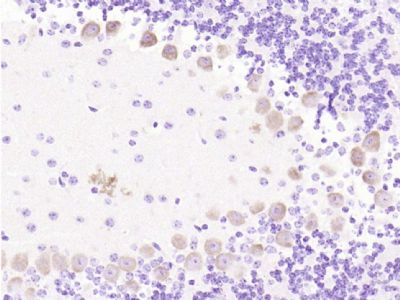 Paraformaldehyde-fixed, paraffin embedded (mouse cerebellum); Antigen retrieval by boiling in sodium citrate buffer (pH6.0) for 15min; Block endogenous peroxidase by 3% hydrogen peroxide for 20 minutes; Blocking buffer (normal goat serum) at 37°C for 30min; Antibody incubation with (phospho-GAP43 (Ser41)) Polyclonal Antibody, Unconjugated (bs-1641R) at 1:200 overnight at 4°C, followed by operating according to SP Kit(Rabbit) (sp-0023) instructionsand DAB staining. Paraformaldehyde-fixed, paraffin embedded (mouse cerebellum); Antigen retrieval by boiling in sodium citrate buffer (pH6.0) for 15min; Block endogenous peroxidase by 3% hydrogen peroxide for 20 minutes; Blocking buffer (normal goat serum) at 37°C for 30min; Antibody incubation with (phospho-GAP43 (Ser41)) Polyclonal Antibody, Unconjugated (bs-1641R) at 1:200 overnight at 4°C, followed by operating according to SP Kit(Rabbit) (sp-0023) instructionsand DAB staining.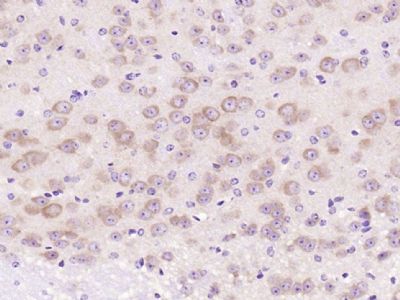 Paraformaldehyde-fixed, paraffin embedded (mouse brain); Antigen retrieval by boiling in sodium citrate buffer (pH6.0) for 15min; Block endogenous peroxidase by 3% hydrogen peroxide for 20 minutes; Blocking buffer (normal goat serum) at 37°C for 30min; Antibody incubation with (phospho-GAP43 (Ser41)) Polyclonal Antibody, Unconjugated (bs-1641R) at 1:200 overnight at 4°C, followed by operating according to SP Kit(Rabbit) (sp-0023) instructionsand DAB staining. Paraformaldehyde-fixed, paraffin embedded (mouse brain); Antigen retrieval by boiling in sodium citrate buffer (pH6.0) for 15min; Block endogenous peroxidase by 3% hydrogen peroxide for 20 minutes; Blocking buffer (normal goat serum) at 37°C for 30min; Antibody incubation with (phospho-GAP43 (Ser41)) Polyclonal Antibody, Unconjugated (bs-1641R) at 1:200 overnight at 4°C, followed by operating according to SP Kit(Rabbit) (sp-0023) instructionsand DAB staining.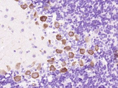 Paraformaldehyde-fixed, paraffin embedded (rat cerebellum); Antigen retrieval by boiling in sodium citrate buffer (pH6.0) for 15min; Block endogenous peroxidase by 3% hydrogen peroxide for 20 minutes; Blocking buffer (normal goat serum) at 37°C for 30min; Antibody incubation with (phospho-GAP43 (Ser41)) Polyclonal Antibody, Unconjugated (bs-1641R) at 1:200 overnight at 4°C, followed by operating according to SP Kit(Rabbit) (sp-0023) instructionsand DAB staining. Paraformaldehyde-fixed, paraffin embedded (rat cerebellum); Antigen retrieval by boiling in sodium citrate buffer (pH6.0) for 15min; Block endogenous peroxidase by 3% hydrogen peroxide for 20 minutes; Blocking buffer (normal goat serum) at 37°C for 30min; Antibody incubation with (phospho-GAP43 (Ser41)) Polyclonal Antibody, Unconjugated (bs-1641R) at 1:200 overnight at 4°C, followed by operating according to SP Kit(Rabbit) (sp-0023) instructionsand DAB staining.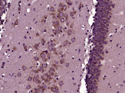 Paraformaldehyde-fixed, paraffin embedded (Rat brain); Antigen retrieval by boiling in sodium citrate buffer (pH6.0) for 15min; Block endogenous peroxidase by 3% hydrogen peroxide for 20 minutes; Blocking buffer (normal goat serum) at 37°C for 30min; Antibody incubation with (phospho-GAP43 (Ser41)) Polyclonal Antibody, Unconjugated (bs-1641R) at 1:400 overnight at 4°C, followed by operating according to SP Kit(Rabbit) (sp-0023) instructionsand DAB staining. Paraformaldehyde-fixed, paraffin embedded (Rat brain); Antigen retrieval by boiling in sodium citrate buffer (pH6.0) for 15min; Block endogenous peroxidase by 3% hydrogen peroxide for 20 minutes; Blocking buffer (normal goat serum) at 37°C for 30min; Antibody incubation with (phospho-GAP43 (Ser41)) Polyclonal Antibody, Unconjugated (bs-1641R) at 1:400 overnight at 4°C, followed by operating according to SP Kit(Rabbit) (sp-0023) instructionsand DAB staining.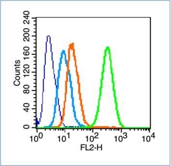 Blank control (blue line): Hela cells (blue). Blank control (blue line): Hela cells (blue).Primary Antibody (green line): Rabbit Anti-phospho-GAP43 (Ser41) antibody (bs-1641R) Dilution: 0.2μg /10^6 cells; Isotype Control Antibody (orange line): Rabbit IgG . Secondary Antibody (white blue line): Goat anti-rabbit IgG-PE Dilution: 1μg /test. Protocol The cells were fixed with 70% methanol (Overnight at 4℃) and then permeabilized with 90% ice-cold methanol for 20 min at -20℃. Cells stained with Primary Antibody for 30 min at room temperature. The cells were then incubated in 1 X PBS/2%BSA/10% goat serum to block non-specific protein-protein interactions followed by the antibody for 15 min at room temperature. The secondary antibody used for 40 min at room temperature. Acquisition of 20,000 events was performed. |


