上海细胞库
人源细胞系| 稳转细胞系| 基因敲除株| 基因点突变细胞株| 基因过表达细胞株| 重组细胞系| 猪的细胞系| 马细胞系| 兔的细胞系| 犬的细胞系| 山羊的细胞系| 鱼的细胞系| 猴的细胞系| 仓鼠的细胞系| 狗的细胞系| 牛的细胞| 大鼠细胞系| 小鼠细胞系| 其他细胞系|

| 规格 | 价格 | 库存 |
|---|---|---|
| 100ul | ¥ 1980 | 200 |
| 产品类型 | 磷酸化抗体 |
| 研究领域 | 免疫学 染色质和核信号 信号转导 转录调节因子 |
| 抗体来源 | Rabbit |
| 克隆类型 | Polyclonal |
| 交叉反应 | Human, Mouse, Rat, (predicted: Chicken, Dog, Pig, Cow, Sheep, ) |
| 产品应用 | WB=1:500-2000 ELISA=1:500-1000 IP=1:20-100 IHC-P=1:100-500 IHC-F=1:100-500 Flow-Cyt=1μg/Test IF=1:100-500 (石蜡切片需做抗原修复) not yet tested in other applications. optimal dilutions/concentrations should be determined by the end user. |
| 分 子 量 | 37kDa |
| 细胞定位 | 细胞核 |
| 性 状 | Liquid |
| 浓 度 | 1mg/ml |
| 免 疫 原 | KLH conjugated Synthesised phosphopeptide derived from human CREB-1 around the phosphorylation site of Ser133:RP(p-S)YR |
| 亚 型 | IgG |
| 纯化方法 | affinity purified by Protein A |
| 储 存 液 | 0.01M TBS(pH7.4) with 1% BSA, 0.03% Proclin300 and 50% Glycerol. |
| 保存条件 | Shipped at 4℃. Store at -20 °C for one year. Avoid repeated freeze/thaw cycles. |
| PubMed | PubMed |
| 产品介绍 | The ATF/CREB family consists of transcription factors that function through binding to the cAMP responsive element (CRE) palindromic octanucleotide, TGACCTCA. The best characterized members of this gene family include CREB-1, CREB-2, ATF-1,ATF-2,ATF-3and ATF-4. these transcription factors share highly-related COOH terminal leucine zipper demerization and basic DNA bindings but are highly divergent in their amino terminal domains. Although each of the ATF/CREB proteins bind CREs in their homodimeric form, in cerain instances they also bind as heterodimers, both within the ATF/CREB family and with members of the AP-1 transcription factor family. It has recentlybeen shown that protein kinase A-mediated CREB phosphorylation results in its binding to a 265kDa nuclear protein designated CBP (CREB-binding protein), which may reprecent a CREB co-activator. Function: Phosphorylation-dependent transcription factor that stimulates transcription upon binding to the DNA cAMP response element (CRE), a sequence present in many viral and cellular promoters. Transcription activation is enhanced by the TORC coactivators which act independently of Ser-133 phosphorylation. Involved in different cellular processes including the synchronization of circadian rhythmicity and the differentiation of adipose cells.independently of Ser-133 phosphorylation. Involved in different cellular processes including the synchronization of circadian rhythmicity and the differentiation of adipose cells. [SUBUNIT] Interacts with PPRC1. Binds DNA as a dimer. This dimer is stabilized by magnesium ions. Interacts, through the bZIP domain, with the coactivators TORC1/CRTC1, TORC2/CRTC2 and TORC3/CRTC3. When phosphorylated on Ser-133, binds CREBBP (By similarity). Interacts with CREBL2; regulates CREB1 phosphorylation, stability and transcriptional activity (By similarity). Interacts (phosphorylated form) with TOX3. Interacts with ARRB1. Binds to HIPK2. Interacts with SGK1. Subunit: Interacts with PPRC1. Binds DNA as a dimer. This dimer is stabilized by magnesium ions. Interacts, through the bZIP domain, with the coactivators TORC1/CRTC1, TORC2/CRTC2 and TORC3/CRTC3. When phosphorylated on Ser-133, binds CREBBP (By similarity). Interacts with CREBL2; regulates CREB1 phosphorylation, stability and transcriptional activity (By similarity). Interacts (phosphorylated form) with TOX3. Interacts with ARRB1. Binds to HIPK2. Interacts with SGK1. Subcellular Location: Nucleus. Post-translational modifications: Stimulated by phosphorylation. Phosphorylation of both Ser-133 and Ser-142 in the SCN regulates the activity of CREB and participates in circadian rhythm generation. Phosphorylation of Ser-133 allows CREBBP binding (By similarity). CREBL2 positively regulates phosphorylation at Ser-133 thereby stimulating CREB1 transcriptional activity (By similarity). Phosphorylated upon DNA damage, probably by ATM or ATR. Phosphorylated upon calcium influx by CaMK4 and CaMK2 on Ser-133. CaMK4 is much more potent than CaMK2 in activating CREB. Phosphorylated by CaMK2 on Ser-142. Phosphorylation of Ser-142 blocks CREB-mediated transcription even when Ser-133 is phosphorylated. Phosphorylated by CaMK1 (By similarity). Phosphorylation of Ser-271 by HIPK2 in response to genotoxic stress promotes CREB1 activity, facilitating the recruitment of the coactivator CBP. Phosphorylated at Ser-133 by RPS6KA3, RPS6KA4 and RPS6KA5 in response to mitogenic or stress stimuli. Sumoylated with SUMO1. Sumoylation on Lys-304, but not on Lys-285, is required for nuclear localization of this protein. Sumoylation is enhanced under hypoxia, promoting nuclear localization and stabilization. DISEASE: Defects in CREB1 may be a cause of angiomatoid fibrous histiocytoma (AFH) [MIM:612160]. A distinct variant of malignant fibrous histiocytoma that typically occurs in children and adolescents and is manifest by nodular subcutaneous growth. Characteristic microscopic features include lobulated sheets of histiocyte-like cells intimately associated with areas of hemorrhage and cystic pseudovascular spaces, as well as a striking cuffing of inflammatory cells, mimicking a lymph node metastasis. Note=A chromosomal aberration involving CREB1 is found in a patient with angiomatoid fibrous histiocytoma. Translocation t(2;22)(q33;q12) with CREB1 generates a EWSR1/CREB1 fusion gene that is most common genetic abnormality in this tumor type. Note=A CREB1 mutation has been found in a patient with multiple congenital anomalies consisting of agenesis of the corpus callosum, cerebellar hypoplasia, severe neonatal respiratory distress refractory to surfactant, thymus hypoplasia, and thyroid follicular hypoplasia (PubMed:22267179). Similarity: Belongs to the bZIP family. Contains 1 bZIP domain. Contains 1 KID (kinase-inducible) domain. SWISS: P16220 Gene ID: 1385 Database links: Entrez Gene: 281713 Cow Entrez Gene: 1385 Human Entrez Gene: 12912 Mouse Entrez Gene: 81646 Rat Omim: 123810 Human SwissProt: P27925 Cow SwissProt: P16220 Human SwissProt: Q01147 Mouse SwissProt: P15337 Rat Unigene: 516646 Human Unigene: 453295 Mouse Unigene: 90061 Rat Important Note: This product as supplied is intended for research use only, not for use in human, therapeutic or diagnostic applications. 磷酸化环腺苷酸应答元件结合蛋白(p-cAMP responsive element binding protein-1, p-CREB-1)是真核细胞转录因子,属于ATF/CREB家族。参与由cAMP或某些病毒蛋白质所诱导基因转录的调节。 |
| 产品图片 | 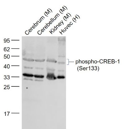 Sample: Sample:Lane 1: Cerebrum (Mouse) Lysate at 40 ug Lane 2: Cerebellum (Mouse) Lysate at 40 ug Lane 3: Kidney (Mouse) Lysate at 40 ug Lane 4: Huvec (Human) Cell Lysate at 30 ug Primary: Anti-phospho-CREB-1 (Ser133) (bs-0036R) at 1/1000 dilution Secondary: IRDye800CW Goat Anti-Rabbit IgG at 1/20000 dilution Predicted band size: 43 kD Observed band size: 45 kD 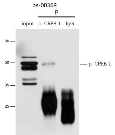 P-CREB1 was immunoprecipitated from mouse kidney tissue with bs-0036R at 1/150 dilution. Western blot was performed from the immunoprecipitate using protein A/G beads. HRP Conjugated Mouse anti-Rabbit IgG (Light Chain specific) was used as secondary antibody at 1:5000 dilution. P-CREB1 was immunoprecipitated from mouse kidney tissue with bs-0036R at 1/150 dilution. Western blot was performed from the immunoprecipitate using protein A/G beads. HRP Conjugated Mouse anti-Rabbit IgG (Light Chain specific) was used as secondary antibody at 1:5000 dilution.Lane 1: mouse kidney tissue lysate 10 µg (Input). Lane 2: bs-0036R IP in mouse kidney tissue lysate. Lane 3: native rabbit IgG IP in mouse kidney tissue lysate (negative control). Secondary All lanes : Mouse anti-Rabbit IgG (Light Chain specific), HRP Conjugated, 1:5000 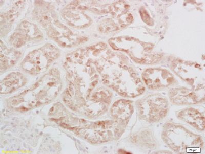 Tissue/cell:bs-0036R human kidney tissue; 4% Paraformaldehyde-fixed and paraffin-embedded; Tissue/cell:bs-0036R human kidney tissue; 4% Paraformaldehyde-fixed and paraffin-embedded;Antigen retrieval: citrate buffer ( 0.01M, pH 6.0 ), Boiling bathing for 15min; Block endogenous peroxidase by 3% Hydrogen peroxide for 30min; Blocking buffer (normal goat serum,C-0005) at 37℃ for 20 min; Incubation: Anti-phospho-CREB-1(Ser133) Polyclonal Antibody, Unconjugated(bs-0036R) 1:200, overnight at 4°C, followed by conjugation to the secondary antibody(SP-0023) and DAB(C-0010) staining 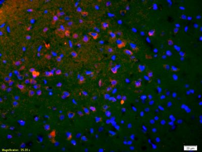 Tissue/cell: rat brain tissue;4% Paraformaldehyde-fixed and paraffin-embedded; Tissue/cell: rat brain tissue;4% Paraformaldehyde-fixed and paraffin-embedded;Antigen retrieval: citrate buffer ( 0.01M, pH 6.0 ), Boiling bathing for 15min; Blocking buffer (normal goat serum,C-0005) at 37℃ for 20 min; Incubation: Anti-phospho-CREB-1(Ser133) Polyclonal Antibody, Unconjugated(bs-0036R) 1:200, overnight at 4°C; The secondary antibody was Goat Anti-Rabbit IgG, Cy3 conjugated (bs-0295G-Cy3)used at 1:200 dilution for 40 minutes at 37°C. DAPI(5ug/ml,blue,C-0033) was used to stain the cell nuclei 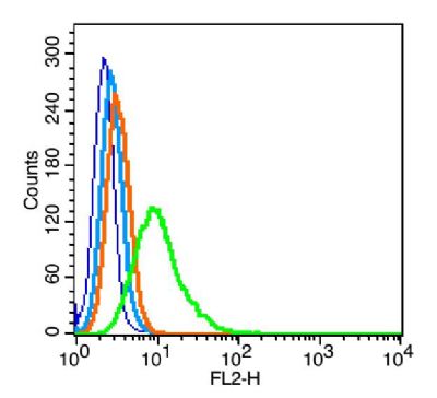 Blank control: RSC96(blue), the cells were fixed with 2% paraformaldehyde (10 min) and then permeabilized with ice-cold 90% methanol for 30 min on ice. Blank control: RSC96(blue), the cells were fixed with 2% paraformaldehyde (10 min) and then permeabilized with ice-cold 90% methanol for 30 min on ice.Isotype Control Antibody: Rabbit IgG(orange) ; Secondary Antibody: Goat anti-rabbit IgG-PE(white blue), Dilution: 1:200 in 1 X PBS containing 0.5% BSA ; Primary Antibody Dilution: 1μg in 100 μL1X PBS containing 0.5% BSA(green). |