上海细胞库
人源细胞系| 稳转细胞系| 基因敲除株| 基因点突变细胞株| 基因过表达细胞株| 重组细胞系| 猪的细胞系| 马细胞系| 兔的细胞系| 犬的细胞系| 山羊的细胞系| 鱼的细胞系| 猴的细胞系| 仓鼠的细胞系| 狗的细胞系| 牛的细胞| 大鼠细胞系| 小鼠细胞系| 其他细胞系|

| 规格 | 价格 | 库存 |
|---|---|---|
| 50ul | ¥ 980 | 200 |
| 100ul | ¥ 1680 | 200 |
| 200ul | ¥ 2480 | 200 |
| 中文名称 | B7-H4抗体 |
| 别 名 | B7-H4; B7h4; B7S1; B7x; BC032925; Immune costimulatory protein B7H4; MGC41287; PRO1291; T cell costimulatory molecule B7x; V set domain-containing T cell activation inhibitor 1; VCTN1; VTCN1_HUMAN. |
| 研究领域 | 肿瘤 免疫学 转录调节因子 |
| 抗体来源 | Rabbit |
| 克隆类型 | Polyclonal |
| 交叉反应 | Human, (predicted: Mouse, Rat, Dog, ) |
| 产品应用 | ELISA=1:500-1000 IHC-P=1:100-500 IHC-F=1:100-500 Flow-Cyt=1μg /test IF=1:100-500 (石蜡切片需做抗原修复) not yet tested in other applications. optimal dilutions/concentrations should be determined by the end user. |
| 分 子 量 | 28kDa |
| 细胞定位 | 细胞膜 |
| 性 状 | Liquid |
| 浓 度 | 1mg/ml |
| 免 疫 原 | KLH conjugated synthetic peptide derived from human B7H4:50-100/282 |
| 亚 型 | IgG |
| 纯化方法 | affinity purified by Protein A |
| 储 存 液 | 0.01M TBS(pH7.4) with 1% BSA, 0.03% Proclin300 and 50% Glycerol. |
| 保存条件 | Shipped at 4℃. Store at -20 °C for one year. Avoid repeated freeze/thaw cycles. |
| PubMed | PubMed |
| 产品介绍 | B7-H4 protein is expressed on the surface of a variety of immune cells and functions as a negative regulator of T cell responses. While B7-H4 mRNA is widely distributed in mouse and human peripheral tissues, cell surface expression of B7-H4 protein is limited and shows an inducible pattern on hematopoietic cells. Putative receptor of B7-H4 can be upregulated on activated T cells. By arresting cell cycle, B7-H4 ligation of T cells has a profound inhibitory effect on the growth, cytokine secretion, and development of cytotoxicity. Administration of B7-H4Ig into mice impairs antigen-specific T cell responses whereas blockade of endogenous B7-H4 by specific monoclonal antibody promotes T cell responses. B7-H4 thus may participate in negative regulation of cell-mediated immunity in peripheral tissues. Function: Negatively regulates T-cell-mediated immune response by inhibiting T-cell activation, proliferation, cytokine production and development of cytotoxicity. When expressed on the cell surface of tumor macrophages, plays an important role, together with regulatory T-cells (Treg), in the suppression of tumor-associated antigen-specific T-cell immunity. Involved in promoting epithelial cell transformation. Subcellular Location: cell membrane; Single-pass type I membrane protein (Potential). Note=Expressed at the cell surface. A soluble form has also been detected. Tissue Specificity: Overexpressed in breast, ovarian, endometrial, renal cell (RCC) and non-small-cell lung cancers (NSCLC). Expressed on activated T- and B-cells, monocytes and dendritic cells, but not expressed in most normal tissues (at protein level). Widely expressed, including in kidney, liver, lung, ovary, placenta, spleen and testis. Post-translational modifications: N-glycosylated. Similarity: Belongs to the immunoglobulin superfamily. BTN/MOG family. Contains 2 Ig-like V-type (immunoglobulin-like) domains. SWISS: Q501W4 Gene ID: 79679 Database links: Entrez Gene: 79679 Human Entrez Gene: 242122 Mouse Entrez Gene: 295322 Rat Omim: 608162 Human SwissProt: Q7Z7D3 Human SwissProt: Q7TSP5 Mouse SwissProt: Q501W4 Rat Unigene: 546434 Human Unigene: 137467 Mouse Unigene: 160956 Rat Important Note: This product as supplied is intended for research use only, not for use in human, therapeutic or diagnostic applications. B7-H4(B7 Homolog 4)是B7家族中的新成员,它能通过抑制T细胞的增殖、细胞因子的产生和细胞周期的进程来负性调控T细胞的免疫应答,其大量表达B7-H4还可以促进上皮细胞的恶性转化,保护表皮细胞免于失巢凋亡,在肿瘤的发生,进展和转归中发挥重要作用. 目前对B7-H4信号通路的进一步的研究必将为自身免疫性疾病、病毒感染性疾病和器官移植后排斥反应中T细胞介导的免疫应答调控提供了新的途径,同时也为肿瘤的诊断、治疗提供崭新的前景。 |
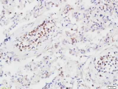
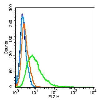
| 产品图片 | Tissue/cell: human colon carcinoma; 4% Paraformaldehyde-fixed and paraffin-embedded; Antigen retrieval: citrate buffer ( 0.01M, pH 6.0 ), Boiling bathing for 15min; Block endogenous peroxidase by 3% Hydrogen peroxide for 30min; Blocking buffer (normal goat serum,C-0005) at 37℃ for 20 min; Incubation: Anti-B7H4 Polyclonal Antibody, Unconjugated(bs-0673R) 1:200, overnight at 4°C, followed by conjugation to the secondary antibody(SP-0023) and DAB(C-0010) staining  Paraformaldehyde-fixed, paraffin embedded (Human lung cancer); Antigen retrieval by boiling in sodium citrate buffer (pH6.0) for 15min; Block endogenous peroxidase by 3% hydrogen peroxide for 20 minutes; Blocking buffer (normal goat serum) at 37°C for 30min; Antibody incubation with (B7H4) Polyclonal Antibody, Unconjugated (bs-0673R) at 1:400 overnight at 4°C, followed by operating according to SP Kit(Rabbit) (sp-0023) instructions and DAB staining.Tissue/cell: human gastric carcinoma; 4% Paraformaldehyde-fixed and paraffin-embedded; Paraformaldehyde-fixed, paraffin embedded (Human lung cancer); Antigen retrieval by boiling in sodium citrate buffer (pH6.0) for 15min; Block endogenous peroxidase by 3% hydrogen peroxide for 20 minutes; Blocking buffer (normal goat serum) at 37°C for 30min; Antibody incubation with (B7H4) Polyclonal Antibody, Unconjugated (bs-0673R) at 1:400 overnight at 4°C, followed by operating according to SP Kit(Rabbit) (sp-0023) instructions and DAB staining.Tissue/cell: human gastric carcinoma; 4% Paraformaldehyde-fixed and paraffin-embedded;Antigen retrieval: citrate buffer ( 0.01M, pH 6.0 ), Boiling bathing for 15min; Block endogenous peroxidase by 3% Hydrogen peroxide for 30min; Blocking buffer (normal goat serum,C-0005) at 37℃ for 20 min; Incubation: Anti-B7H4 Polyclonal Antibody, Unconjugated(bs-0673R) 1:200, overnight at 4°C, followed by conjugation to the secondary antibody(SP-0023) and DAB(C-0010) staining Tissue/cell: human thyroid carcinoma; 4% Paraformaldehyde-fixed and paraffin-embedded; Antigen retrieval: citrate buffer ( 0.01M, pH 6.0 ), Boiling bathing for 15min; Block endogenous peroxidase by 3% Hydrogen peroxide for 30min; Blocking buffer (normal goat serum,C-0005) at 37℃ for 20 min; Incubation: Anti-B7H4 Polyclonal Antibody, Unconjugated(bs-0673R) 1:200, overnight at 4°C, followed by conjugation to the secondary antibody(SP-0023) and DAB(C-0010) staining  Blank control: Hela(blue). Blank control: Hela(blue).Primary Antibody:Rabbit Anti-B7H4 antibody(bs-0673R), Dilution: 1μg in 100 μL 1X PBS containing 0.5% BSA; Isotype Control Antibody: Rabbit IgG(orange) ,used under the same conditions ); Secondary Antibody: Goat anti-rabbit IgG-PE(white blue), Dilution: 1:200 in 1 X PBS containing 0.5% BSA. Protocol The cells were fixed with 2% paraformaldehyde (10 min). Antibody (bs-0673R, 1μg /1x10^6 cells) were incubated for 30 min on the ice, followed by 1 X PBS containing 0.5% BSA + 1 0% goat serum (15 min) to block non-specific protein-protein interactions. Then the Goat Anti-rabbit IgG/PE antibody was added into the blocking buffer mentioned above to react with the primary antibody of bs-0673R at 1/200 dilution for 30 min on ice. Acquisition of 20,000 events was performed. |