
上海细胞库
人源细胞系| 稳转细胞系| 基因敲除株| 基因点突变细胞株| 基因过表达细胞株| 重组细胞系| 猪的细胞系| 马细胞系| 兔的细胞系| 犬的细胞系| 山羊的细胞系| 鱼的细胞系| 猴的细胞系| 仓鼠的细胞系| 狗的细胞系| 牛的细胞| 大鼠细胞系| 小鼠细胞系| 其他细胞系|
人源细胞系| 稳转细胞系| 基因敲除株| 基因点突变细胞株| 基因过表达细胞株| 重组细胞系| 猪的细胞系| 马细胞系| 兔的细胞系| 犬的细胞系| 山羊的细胞系| 鱼的细胞系| 猴的细胞系| 仓鼠的细胞系| 狗的细胞系| 牛的细胞| 大鼠细胞系| 小鼠细胞系| 其他细胞系|

| 规格 | 价格 | 库存 |
|---|---|---|
| 50ul | ¥ 980 | 200 |
| 100ul | ¥ 1680 | 200 |
| 200ul | ¥ 2200 | 200 |
| 中文名称 | 干扰素诱导蛋白AIM2抗体 |
| 别 名 | Absent in melanoma 2; AIM 2; Aim2; AIM2_MOUSE; Interferon-inducible protein AIM2; OTTHUMP00000035296; PYHIN4. |
| 研究领域 | 肿瘤 免疫学 染色质和核信号 转录调节因子 表观遗传学 |
| 抗体来源 | Rabbit |
| 克隆类型 | Polyclonal |
| 交叉反应 | Human, Mouse, Rat, |
| 产品应用 | WB=1:500-2000 ELISA=1:500-1000 IHC-P=1:100-500 IHC-F=1:100-500 Flow-Cyt=2μg/Test IF=1:100-500 (石蜡切片需做抗原修复) not yet tested in other applications. optimal dilutions/concentrations should be determined by the end user. |
| 分 子 量 | 40kDa |
| 细胞定位 | 细胞核 细胞浆 |
| 性 状 | Liquid |
| 浓 度 | 1mg/ml |
| 免 疫 原 | KLH conjugated synthetic peptide derived from mouse AIM2.:255-354/354 |
| 亚 型 | IgG |
| 纯化方法 | affinity purified by Protein A |
| 储 存 液 | 0.01M TBS(pH7.4) with 1% BSA, 0.03% Proclin300 and 50% Glycerol. |
| 保存条件 | Shipped at 4℃. Store at -20 °C for one year. Avoid repeated freeze/thaw cycles. |
| PubMed | PubMed |
| 产品介绍 | AIM2 is a member of the IFI20X /IFI16 family. It plays a putative role in tumorigenic reversion and may control cell proliferation. Interferon-gamma induces expression of AIM2. Function: Involved in innate immune response by recognizing cytosolic double-stranded DNA and inducing caspase-1-activating inflammasome formation in macrophages. Upon binding to DNA is thought to undergo oligomerization and to associate with PYCARD initiating the recruitment of caspase-1 precusrsor and processing of interleukin-1 beta and interleukin-18. Detects cytosolic dsDNA of viral and bacterial origin in a non-sequence-specific manner. Can also trigger PYCARD-dependent, caspase-1-independent cell death that involves caspase-8. Subunit: Self-associates; forms homooligomers in response to cytosolic dsDNA and the dsDNA seems to serve as oligomerization platform. Component of the AIM2 inflammasome complex. Interacts with PYCARD. Interacts with IFI16 (By similarity). Interacts with EIF2AK2/PKR (By similarity). Subcellular Location: Nucleus (By similarity). Cytoplasm. Tissue Specificity: Expressed in spleen, small intestine, peripheral blood leukocytes, and testis. Similarity: Belongs to the HIN-200 family. Contains 1 DAPIN domain. Contains 1 HIN-200 domain. SWISS: Q91VJ1 Gene ID: 383619 Database links: Entrez Gene: 9447 Human Entrez Gene: 383619 Mouse Entrez Gene: 304987 Rat Omim: 604578 Human SwissProt: O14862 Human SwissProt: Q91VJ1 Mouse Unigene: 281898 Human Unigene: 733411 Human Unigene: 131453 Mouse Important Note: This product as supplied is intended for research use only, not for use in human, therapeutic or diagnostic applications. |
| 产品图片 | 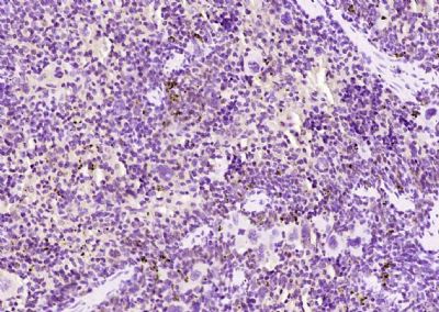 Paraformaldehyde-fixed, paraffin embedded (mouse spleen); Antigen retrieval by boiling in sodium citrate buffer (pH6.0) for 15min; Block endogenous peroxidase by 3% hydrogen peroxide for 20 minutes; Blocking buffer (normal goat serum) at 37°C for 30min; Antibody incubation with (AIM2) Polyclonal Antibody, Unconjugated (bs-5986R) at 1:200 overnight at 4°C, followed by operating according to SP Kit(Rabbit) (sp-0023) instructionsand DAB staining. Paraformaldehyde-fixed, paraffin embedded (mouse spleen); Antigen retrieval by boiling in sodium citrate buffer (pH6.0) for 15min; Block endogenous peroxidase by 3% hydrogen peroxide for 20 minutes; Blocking buffer (normal goat serum) at 37°C for 30min; Antibody incubation with (AIM2) Polyclonal Antibody, Unconjugated (bs-5986R) at 1:200 overnight at 4°C, followed by operating according to SP Kit(Rabbit) (sp-0023) instructionsand DAB staining.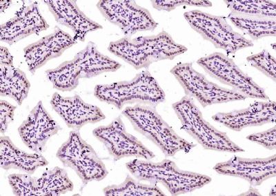 Paraformaldehyde-fixed, paraffin embedded (mouse intestine); Antigen retrieval by boiling in sodium citrate buffer (pH6.0) for 15min; Block endogenous peroxidase by 3% hydrogen peroxide for 20 minutes; Blocking buffer (normal goat serum) at 37°C for 30min; Antibody incubation with (AIM2) Polyclonal Antibody, Unconjugated (bs-5986R) at 1:200 overnight at 4°C, followed by operating according to SP Kit(Rabbit) (sp-0023) instructionsand DAB staining. Paraformaldehyde-fixed, paraffin embedded (mouse intestine); Antigen retrieval by boiling in sodium citrate buffer (pH6.0) for 15min; Block endogenous peroxidase by 3% hydrogen peroxide for 20 minutes; Blocking buffer (normal goat serum) at 37°C for 30min; Antibody incubation with (AIM2) Polyclonal Antibody, Unconjugated (bs-5986R) at 1:200 overnight at 4°C, followed by operating according to SP Kit(Rabbit) (sp-0023) instructionsand DAB staining.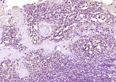 Paraformaldehyde-fixed, paraffin embedded (human tonsil); Antigen retrieval by boiling in sodium citrate buffer (pH6.0) for 15min; Block endogenous peroxidase by 3% hydrogen peroxide for 20 minutes; Blocking buffer (normal goat serum) at 37°C for 30min; Antibody incubation with (AIM2) Polyclonal Antibody, Unconjugated (bs-5986R) at 1:200 overnight at 4°C, followed by operating according to SP Kit(Rabbit) (sp-0023) instructionsand DAB staining. Paraformaldehyde-fixed, paraffin embedded (human tonsil); Antigen retrieval by boiling in sodium citrate buffer (pH6.0) for 15min; Block endogenous peroxidase by 3% hydrogen peroxide for 20 minutes; Blocking buffer (normal goat serum) at 37°C for 30min; Antibody incubation with (AIM2) Polyclonal Antibody, Unconjugated (bs-5986R) at 1:200 overnight at 4°C, followed by operating according to SP Kit(Rabbit) (sp-0023) instructionsand DAB staining.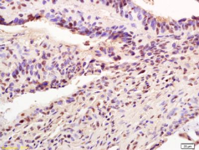 Tissue/cell: human colon carcinoma; 4% Paraformaldehyde-fixed and paraffin-embedded; Tissue/cell: human colon carcinoma; 4% Paraformaldehyde-fixed and paraffin-embedded;Antigen retrieval: citrate buffer ( 0.01M, pH 6.0 ), Boiling bathing for 15min; Block endogenous peroxidase by 3% Hydrogen peroxide for 30min; Blocking buffer (normal goat serum,C-0005) at 37℃ for 20 min; Incubation: Anti-AIM2 Polyclonal Antibody, Unconjugated(bs-5986R) 1:200, overnight at 4°C, followed by conjugation to the secondary antibody(SP-0023) and DAB(C-0010) staining Image was kindly submitted by a researcher at Duke University Medical Center. Rat splenocytes stained with Rabbit Anti-AIM2 Polyclonal Antibody, PE conjugated (bs-5986R-PE)at 1:25. 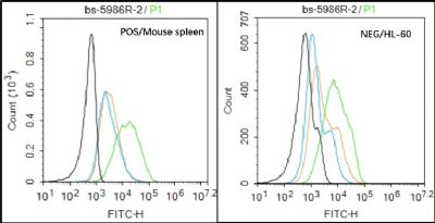 Black line : Positive blank control (Mouse spleen); Negative blank control (HL60) Black line : Positive blank control (Mouse spleen); Negative blank control (HL60)Green line : Primary Antibody (Rabbit Anti-AIM2 antibody (bs-5986R) ) Orange line:Isotype Control Antibody (Rabbit IgG) . Blue line : Secondary Antibody (Goat anti-rabbit IgG-AF488) Mouse spleen(Positive)and HL60(Negative control)cells (black) were fixed with 4% PFA for 10min at room temperature, permeabilized with PBST for 20 min at room temperature, and incubated in 5% BSA blocking buffer for 30 min at room temperature. Cells were then stained with AIM2 Antibody(bs-5986R)at 1:50 dilution in blocking buffer and incubated for 30 min at room temperature, washed twice with 2% BSA in PBS, followed by secondary antibody(blue) incubation for 40 min at room temperature. Acquisitions of 20,000 events were performed. Cells stained with primary antibody (green), and isotype control (orange). |


