上海细胞库
人源细胞系| 稳转细胞系| 基因敲除株| 基因点突变细胞株| 基因过表达细胞株| 重组细胞系| 猪的细胞系| 马细胞系| 兔的细胞系| 犬的细胞系| 山羊的细胞系| 鱼的细胞系| 猴的细胞系| 仓鼠的细胞系| 狗的细胞系| 牛的细胞| 大鼠细胞系| 小鼠细胞系| 其他细胞系|

| 规格 | 价格 | 库存 |
|---|---|---|
| 50ul | ¥ 980 | 200 |
| 100ul | ¥ 1680 | 200 |
| 200ul | ¥ 2500 | 200 |
| 中文名称 | 葡萄糖转运蛋白4抗体 |
| 别 名 | insulin-responsive; Glucose transporter GLUT 4; Glucose Transporter GLUT4; Glucose transporter type 4; Glucose transporter type 4 insulin responsive; GLUT 4; GLUT-4; GTR4_HUMAN; kug; SLC 2A4; SLC2A4; solute carrier family 2 (facilitated glucose transporter) member 4; Solute carrier family 2, facilitated glucose transporter member 4. |
| 研究领域 | 肿瘤 心血管 免疫学 信号转导 干细胞 糖尿病 |
| 抗体来源 | Rabbit |
| 克隆类型 | Polyclonal |
| 交叉反应 | Human, Mouse, Rat, (predicted: Dog, Pig, Cow, Rabbit, Sheep, ) |
| 产品应用 | WB=1:500-2000 ELISA=1:500-1000 IHC-P=1:100-500 IHC-F=1:100-500 Flow-Cyt=1μg/Test ICC=1:100 IF=1:100-500 (石蜡切片需做抗原修复) not yet tested in other applications. optimal dilutions/concentrations should be determined by the end user. |
| 分 子 量 | 54kDa |
| 细胞定位 | 细胞浆 细胞膜 |
| 性 状 | Liquid |
| 浓 度 | 1mg/ml |
| 免 疫 原 | KLH conjugated synthetic peptide derived from human GLUT4:401-509/509 |
| 亚 型 | IgG |
| 纯化方法 | affinity purified by Protein A |
| 储 存 液 | 0.01M TBS(pH7.4) with 1% BSA, 0.03% Proclin300 and 50% Glycerol. |
| 保存条件 | Shipped at 4℃. Store at -20 °C for one year. Avoid repeated freeze/thaw cycles. |
| PubMed | PubMed |
| 产品介绍 | GLUT4 is the facilitated glucose transporter expressed exclusively in adipocytes and muscle cells, and is also known as the "insulin-responsive" glucose transporter. GLUT4 translocates from an ill-defined intracellular compartment to the plasma membrane in response to insulin. The total cellular content of GLUT4 is significantly decreased in adipose cells from many patients with Type II diabetes mellitus, and animals with some types of experimental diabetes. Function: Insulin-regulated facilitative glucose transporter. Subcellular Location: Endomembrane system. Cytoplasm > perinuclear region. Localizes primarily to the perinuclear region, undergoing continued recycling to the plasma membrane where it is rapidly reinternalized. The dileucine internalization motif is critical for intracellular sequestration. Tissue Specificity: Skeletal and cardiac muscles; brown and white fat. Post-translational modifications: Sumoylated. DISEASE: Defects in SLC2A4 may be a cause of noninsulin-dependent diabetes mellitus (NIDDM) [MIM:125853]. Defects in SLC2A4 may be a cause of certain post-receptor defects in NIDDM. The variant in position Ile-383 is found in a small number of NIDDM patients, but seems not to be found in nondiabetic subjects. Similarity: Belongs to the major facilitator superfamily. Sugar transporter (TC 2.A.1.1) family. Glucose transporter subfamily. SWISS: P14672 Gene ID: 6517 Database links: Entrez Gene: 282359 Cow Entrez Gene: 6517 Human Entrez Gene: 20528 Mouse Entrez Gene: 25139 Rat Omim: 138190 Human SwissProt: Q27994 Cow SwissProt: Q29RP5 Cow SwissProt: P14672 Human SwissProt: P14142 Mouse SwissProt: P19357 Rat Unigene: 380691 Human Unigene: 10661 Mouse Unigene: 1314 Rat Important Note: This product as supplied is intended for research use only, not for use in human, therapeutic or diagnostic applications. 葡萄糖转运蛋白-4是一种十分重要的葡萄糖转运体,与胰岛素抵抗和2型糖尿病密切相关.葡萄糖转运蛋白4在细胞内部和细胞膜之间循环流动.实现对葡萄糖的转运需要葡萄糖转运蛋白4自身的转位和活化.葡萄糖转运蛋白4与肥胖、肿瘤相关联. GLUT-2和GLUT-4蛋白这两个葡萄糖运载体的研究对于糖尿病的胰岛素释放障碍和胰岛素抵抗有重要意义. |
| 产品图片 | 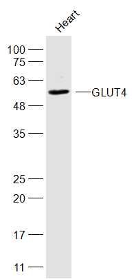 Sample: Sample:Heart(Rat) Cell Lysate at 40 ug Primary: Anti-GLUT4 (bs-0384R) at 1/300 dilution Secondary: IRDye800CW Goat Anti-Rabbit IgG at 1/20000 dilution Predicted band size: 54 kD Observed band size: 54 kD 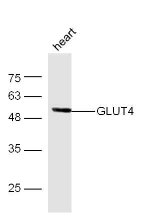 Sample: Heart (Mouse) Lysate at 40 ug Sample: Heart (Mouse) Lysate at 40 ugPrimary: Anti- GLUT4 (bs-0384R) at 1/300 dilution Secondary: IRDye800CW Goat Anti-Rabbit IgG at 1/10000 dilution Predicted band size: 54 kD Observed band size: 54 kD 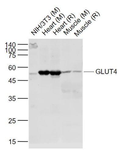 Sample: Sample:Lane 1: NIH/3T3 (Mouse) Cell Lysate at 30 ug Lane 2: Heart (Mouse) Lysate at 40 ug Lane 3: Heart (Rat) Lysate at 40 ug Lane 4: Muscle (Mouse) Lysate at 40 ug Lane 5: Muscle (Rat) Lysate at 40 ug Primary: Anti-GLUT4 (bs-0384R) at 1/1000 dilution Secondary: IRDye800CW Goat Anti-Rabbit IgG at 1/20000 dilution Predicted band size: 51 kD Observed band size: 51 kD 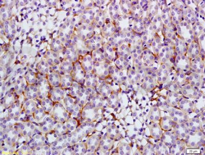 Tissue/cell: rat kidney tissue; 4% Paraformaldehyde-fixed and paraffin-embedded; Tissue/cell: rat kidney tissue; 4% Paraformaldehyde-fixed and paraffin-embedded;Antigen retrieval: citrate buffer ( 0.01M, pH 6.0 ), Boiling bathing for 15min; Block endogenous peroxidase by 3% Hydrogen peroxide for 30min; Blocking buffer (normal goat serum,C-0005) at 37℃ for 20 min; Incubation: Anti-GLUT4 Polyclonal Antibody, Unconjugated(bs-0384R) 1:200, overnight at 4°C, followed by conjugation to the secondary antibody(SP-0023) and DAB(C-0010) staining 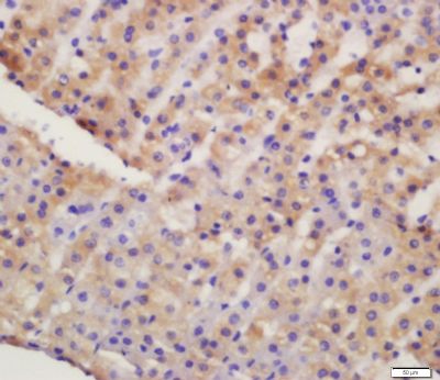 Tissue/cell: rat kidney tissue; 4% Paraformaldehyde-fixed and paraffin-embedded; Tissue/cell: rat kidney tissue; 4% Paraformaldehyde-fixed and paraffin-embedded;Antigen retrieval: citrate buffer ( 0.01M, pH 6.0 ), Boiling bathing for 15min; Block endogenous peroxidase by 3% Hydrogen peroxide for 30min; Blocking buffer (normal goat serum,C-0005) at 37℃ for 20 min; Incubation: Anti-GLUT4 Polyclonal Antibody, Unconjugated(bs-0384R) 1:200, overnight at 4°C, followed by conjugation to the secondary antibody(SP-0023) and DAB(C-0010) staining 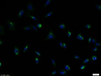 Tissue/cell: NIH/3T3 cell; 4% Paraformaldehyde-fixed; Triton X-100 at room temperature for 20 min; Blocking buffer (normal goat serum, C-0005) at 37°C for 20 min; Antibody incubation with (GLUT4) polyclonal Antibody, Unconjugated (bs-0384R) 1:100, 90 minutes at 37°C; followed by a FITC conjugated Goat Anti-Rabbit IgG antibody at 37°C for 90 minutes, DAPI (blue, C02-04002) was used to stain the cell nuclei. Tissue/cell: NIH/3T3 cell; 4% Paraformaldehyde-fixed; Triton X-100 at room temperature for 20 min; Blocking buffer (normal goat serum, C-0005) at 37°C for 20 min; Antibody incubation with (GLUT4) polyclonal Antibody, Unconjugated (bs-0384R) 1:100, 90 minutes at 37°C; followed by a FITC conjugated Goat Anti-Rabbit IgG antibody at 37°C for 90 minutes, DAPI (blue, C02-04002) was used to stain the cell nuclei.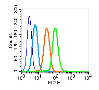 Blank control (blue line): K562 (blue). Blank control (blue line): K562 (blue).Primary Antibody (green line): Rabbit Anti-GLUT4 antibody (bs-0384R) Dilution: 1μg /10^6 cells; Isotype Control Antibody (orange line): Rabbit IgG . Secondary Antibody (white blue line): Goat anti-rabbit IgG-PE Dilution: 1μg /test. Protocol The cells were fixed with 70% ethanol (overnight at 4℃) and then permeabilized with 0.1% PBS-Tween for 20 min at room temperature. Cells stained with Primary Antibody for 30 min at room temperature. The cells were then incubated in 1 X PBS/2%BSA/10% goat serum to block non-specific protein-protein interactions followed by the antibody for 15 min at room temperature. The secondary antibody used for 40 min at room temperature. Acquisition of 20,000 events was performed. |