上海细胞库
人源细胞系| 稳转细胞系| 基因敲除株| 基因点突变细胞株| 基因过表达细胞株| 重组细胞系| 猪的细胞系| 马细胞系| 兔的细胞系| 犬的细胞系| 山羊的细胞系| 鱼的细胞系| 猴的细胞系| 仓鼠的细胞系| 狗的细胞系| 牛的细胞| 大鼠细胞系| 小鼠细胞系| 其他细胞系|

| 规格 | 价格 | 库存 |
|---|---|---|
| 50ul | ¥ 1200 | 200 |
| 100ul | ¥ 1900 | 200 |
| 200ul | ¥ 2900 | 200 |
| 中文名称 | DNA甲基转移酶-3α抗体 |
| 别 名 | DNA (cytosine 5 ) methyltransferase 3 alpha; DNA (cytosine 5) methyltransferase 3A; DNA cytosine methyltransferase 3A2; DNA methyltransferase 3a; DNA methyltransferase HsaIIIA; DNA MTase HsaI; DNA MTase HsaIIIA; DNMT 3a; DNMT; Dnmt3a; DNMT3A2; M HsaIIIA; M.HsaIIIA; MCMT; Methyl CpG binding domain protein 3a. |
| 研究领域 | 肿瘤 细胞生物 信号转导 细胞凋亡 表观遗传学 |
| 抗体来源 | Rabbit |
| 克隆类型 | Polyclonal |
| 交叉反应 | Human, Rat, (predicted: Mouse, Cow, ) |
| 产品应用 | WB=1:500-2000 ELISA=1:500-1000 IHC-P=1:100-500 IHC-F=1:100-500 Flow-Cyt=1ug/Test IF=1:100-500 (石蜡切片需做抗原修复) not yet tested in other applications. optimal dilutions/concentrations should be determined by the end user. |
| 分 子 量 | 100kDa |
| 细胞定位 | 细胞核 细胞浆 |
| 性 状 | Liquid |
| 浓 度 | 1mg/ml |
| 免 疫 原 | KLH conjugated synthetic peptide derived from human Dnmt3a:26-100/912 |
| 亚 型 | IgG |
| 纯化方法 | affinity purified by Protein A |
| 储 存 液 | 0.01M TBS(pH7.4) with 1% BSA, 0.03% Proclin300 and 50% Glycerol. |
| 保存条件 | Shipped at 4℃. Store at -20 °C for one year. Avoid repeated freeze/thaw cycles. |
| PubMed | PubMed |
| 产品介绍 | The tumor organization has the DNA methylation disorder, including and damps cancer gene high methylation DNA with the cell multiplication cycle close related cancer gene low methylation the methyl transferase (Dnmt) participation methylation the formation (is mainly Dnmt3a and Dnmt3b) and the maintenance (is mainly Dnmt1) (isoform CRA_b) (DNA methyltransferase MmuIIIA) (DNA MTase MmuIIIA) Function: Required for genome-wide de novo methylation and is essential for the establishment of DNA methylation patterns during development. DNA methylation is coordinated with methylation of histones. It modifies DNA in a non-processive manner and also methylates non-CpG sites. May preferentially methylate DNA linker between 2 nucleosomal cores and is inhibited by histone H1. Plays a role in paternal and maternal imprinting. Required for methylation of most imprinted loci in germ cells. Acts as a transcriptional corepressor for ZNF238. Can actively repress transcription through the recruitment of HDAC activity. Subunit: Heterotetramer composed of 1 DNMT3A homodimer and 2 DNMT3L subunits (DNMT3L-DNMT3A-DNMT3A-DNMT3L). Interacts with UBC9, PIAS1 and PIAS2. Binds the ZNF238 transcriptional repressor. Interacts with SETDB1. Associates with HDAC1 through its ADD domain. Interacts with DNMT1 and DNMT3B. Interacts with the PRC2/EED-EZH2 complex. Interacts with MPHOSPH8. Interacts with histone H3 that is not methylated at 'Lys-4' (H3K4). Subcellular Location: Nucleus. Cytoplasm. Note=Accumulates in the major satellite repeats at pericentric heterochromatin. Tissue Specificity: Highly expressed in fetal tissues, skeletal muscle, heart, peripheral blood mononuclear cells, kidney, and at lower levels in placenta, brain, liver, colon, spleen, small intestine and lung. Post-translational modifications: Sumoylated; sumoylation disrupts the ability to interact with histone deacetylases (HDAC1 and HDAC2) and repress transcription. Similarity: Belongs to the C5-methyltransferase family. Contains 1 ADD domain. Contains 1 GATA-type zinc finger. Contains 1 PHD-type zinc finger. Contains 1 PWWP domain. SWISS: Q9Y6K1 Gene ID: 1788 Database links: Entrez Gene: 1788 Human Entrez Gene: 13435 Mouse Omim: 602769 Human SwissProt: Q9Y6K1 Human SwissProt: O88508 Mouse Unigene: 515840 Human Unigene: 5001 Mouse Important Note: This product as supplied is intended for research use only, not for use in human, therapeutic or diagnostic applications. 肿瘤组织存在DNA甲基化紊乱,包括与细胞增殖周期密切相关的癌基因低甲基化和抑癌基因高甲基化DNA甲基转移酶(Dnmt)参与甲基化的形成(主要是Dnmt3a和Dnmt3b)和维持(主要是Dnmt1) |
| 产品图片 | 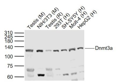 Sample: Sample:Lane 1: Testis (Mouse) Lysate at 40 ug Lane 2: NIH/3T3 (Mouse) Cell Lysate at 30 ug Lane 3: Testis (Rat) Lysate at 40 ug Lane 4: 293T (Human) Cell Lysate at 30 ug Lane 5: SH-SY5Y (Human) Cell Lysate at 30 ug Lane 6: Molt-4 (Human) Cell Lysate at 30 ug Lane 7: HepG2 (Human) Cell Lysate at 30 ug Primary: Anti-Dnmt3a (bs-0497R) at 1/1000 dilution Secondary: IRDye800CW Goat Anti-Rabbit IgG at 1/20000 dilution Predicted band size: 120 kD Observed band size: 120 kD 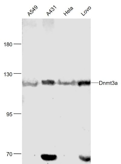 Sample: Sample:A549(Human) Cell Lysate at 30 ug A431(Human) Cell Lysate at 30 ug Hela(Human) Cell Lysate at 30 ug Lovo(Human) Cell Lysate at 30 ug Primary: Anti-Dnmt3a (bs-0497R) at 1/1000 dilution Secondary: IRDye800CW Goat Anti-Rabbit IgG at 1/20000 dilution Predicted band size: 130 kD Observed band size: 120 kD 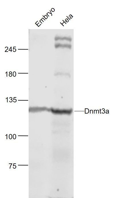 Sample: Sample:Embryo(Mouse) Lysate at 40 ug Hela(Human) Cell Lysate at 30 ug Primary: Anti-Dnmt3a (bs-0497R) at 1/1000 dilution Secondary: IRDye800CW Goat Anti-Rabbit IgG at 1/20000 dilution Predicted band size: 130 kD Observed band size: 120 kD 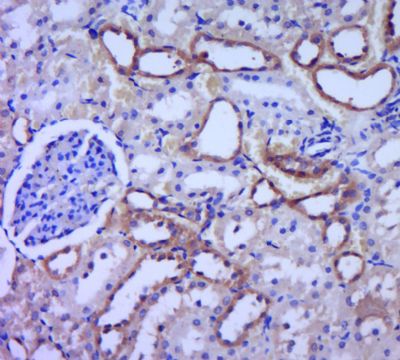 Paraformaldehyde-fixed, paraffin embedded (rat kidney tissue); Antigen retrieval by boiling in sodium citrate buffer (pH6.0) for 15min; Block endogenous peroxidase by 3% hydrogen peroxide for 20 minutes; Blocking buffer (normal goat serum) at 37°C for 30min; Antibody incubation with (Dnmt3a) Polyclonal Antibody, Unconjugated (bs-0497R) at 1:400 overnight at 4°C, followed by a conjugated secondary (sp-0023) for 20 minutes and DAB staining.Tissue/cell: human gastric carcinoma; 4% Paraformaldehyde-fixed and paraffin-embedded; Paraformaldehyde-fixed, paraffin embedded (rat kidney tissue); Antigen retrieval by boiling in sodium citrate buffer (pH6.0) for 15min; Block endogenous peroxidase by 3% hydrogen peroxide for 20 minutes; Blocking buffer (normal goat serum) at 37°C for 30min; Antibody incubation with (Dnmt3a) Polyclonal Antibody, Unconjugated (bs-0497R) at 1:400 overnight at 4°C, followed by a conjugated secondary (sp-0023) for 20 minutes and DAB staining.Tissue/cell: human gastric carcinoma; 4% Paraformaldehyde-fixed and paraffin-embedded;Antigen retrieval: citrate buffer ( 0.01M, pH 6.0 ), Boiling bathing for 15min; Block endogenous peroxidase by 3% Hydrogen peroxide for 30min; Blocking buffer (normal goat serum,C-0005) at 37℃ for 20 min; Incubation: Anti-Dnmt3a Polyclonal Antibody, Unconjugated(bs-0497R) 1:200, overnight at 4°C, followed by conjugation to the secondary antibody(SP-0023) and DAB(C-0010) staining Tissue/cell: human breast carcinoma; 4% Paraformaldehyde-fixed and paraffin-embedded; Antigen retrieval: citrate buffer ( 0.01M, pH 6.0 ), Boiling bathing for 15min; Block endogenous peroxidase by 3% Hydrogen peroxide for 30min; Blocking buffer (normal goat serum,C-0005) at 37℃ for 20 min; Incubation: Anti-Dnmt3a Polyclonal Antibody, Unconjugated(bs-0497R) 1:200, overnight at 4°C, followed by conjugation to the secondary antibody(SP-0023) and DAB(C-0010) staining 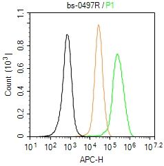 Blank control (Black line): U87MG (Black). Blank control (Black line): U87MG (Black).Primary Antibody (green line): Rabbit Anti-Dnmt3a antibody (bs-0497R) Dilution: 1μg /10^6 cells; Isotype Control Antibody (orange line): Rabbit IgG . Secondary Antibody (white blue line): Goat anti-rabbit IgG-AF647 Dilution: 1μg /test. Protocol The cells were fixed with 4% PFA (10min at room temperature)and then permeabilized with 90% ice-cold methanol for 20 min at room temperature. The cells were then incubated in 5%BSA to block non-specific protein-protein interactions for 30 min at room temperature .Cells stained with Primary Antibody for 30 min at room temperature. The secondary antibody used for 40 min at room temperature. Acquisition of 20,000 events was performed. |