上海细胞库
人源细胞系| 稳转细胞系| 基因敲除株| 基因点突变细胞株| 基因过表达细胞株| 重组细胞系| 猪的细胞系| 马细胞系| 兔的细胞系| 犬的细胞系| 山羊的细胞系| 鱼的细胞系| 猴的细胞系| 仓鼠的细胞系| 狗的细胞系| 牛的细胞| 大鼠细胞系| 小鼠细胞系| 其他细胞系|

| 规格 | 价格 | 库存 |
|---|---|---|
| 50ul | ¥ 1280 | 1 |
| 100ul | ¥ 1980 | 1 |
| 200ul | ¥ 2980 | 1 |
| 中文名称 | CD90抗体 |
| 别 名 | CD90 / Thy1; CD7; CD90 antigen; CDw90; FLJ33325; MGC128895; T25; Theta antigen; Thy-1; Thy 1; Thy 1 cell surface antigen; Thy 1 membrane glycoprotein; Thy 1 membrane glycoprotein precursor; Thy 1.2; Thy-1 T-cell antigen; Thy1 antigen; Thy1 T cell antigen; Thy1.1; Thy1.2; Thymus cell antigen 1, theta; THY1_RAT; THY1_HUMAN. |
| 研究领域 | 细胞生物 免疫学 神经生物学 |
| 抗体来源 | Rabbit |
| 克隆类型 | Polyclonal |
| 交叉反应 | Human, Mouse, Rat, (predicted: Dog, Pig, Horse, Sheep, ) |
| 产品应用 | WB=1:500-2000 IHC-P=1:100-500 IHC-F=1:100-500 Flow-Cyt=1μg/Test IF=1:100-500 (石蜡切片需做抗原修复) not yet tested in other applications. optimal dilutions/concentrations should be determined by the end user. |
| 分 子 量 | 12kDa |
| 细胞定位 | 细胞膜 |
| 性 状 | Liquid |
| 浓 度 | 1mg/ml |
| 免 疫 原 | KLH conjugated synthetic peptide derived from rat Thy-1:31-120/161 |
| 亚 型 | IgG |
| 纯化方法 | affinity purified by Protein A |
| 储 存 液 | 0.01M TBS(pH7.4) with 1% BSA, 0.03% Proclin300 and 50% Glycerol. |
| 保存条件 | Shipped at 4℃. Store at -20 °C for one year. Avoid repeated freeze/thaw cycles. |
| PubMed | PubMed |
| 产品介绍 | Thy-1 or CD90 (Cluster of Differentiation 90) is a 25–37 kDa heavily N-glycosylated, glycophosphatidylinositol (GPI) anchored conserved cell surface protein with a single V-like immunoglobulin domain, originally discovered as a thymocyte antigen. Thy-1can be used as a marker for a variety of stem cells and for the axonal processes of mature neurons. Structural study of Thy-1 lead to the foundation of the Immunoglobulin superfamily, of which it is the smallest member, and led to some of the initial biochemical description and characterization of a vertebrate GPI anchor and also the first demonstration of tissue specific differential glycosylation. Function: May play a role in cell-cell or cell-ligand interactions during synaptogenesis and other events in the brain. Subcellular Location: Cell membrane; Lipid-anchor, GPI-anchor. Tissue Specificity: Abundant in lymphoid tissues. Post-translational modifications: Glycosylation is tissue specific. Sialylation of N-glycans at Asn-93 in brain and at Asn-42, Asn-93 and Asn-117 in thymus. Similarity: Contains 1 Ig-like V-type (immunoglobulin-like) domain. SWISS: P01830 Gene ID: 24832 Database links: Entrez Gene: 7070 Human Entrez Gene: 21838 Mouse Entrez Gene: 24832 Rat Omim: 188230 Human SwissProt: P04216 Human SwissProt: P01831 Mouse SwissProt: P01830 Rat Unigene: 644697 Human Unigene: 3951 Mouse Unigene: 108198 Rat Important Note: This product as supplied is intended for research use only, not for use in human, therapeutic or diagnostic applications. |
| 产品图片 | 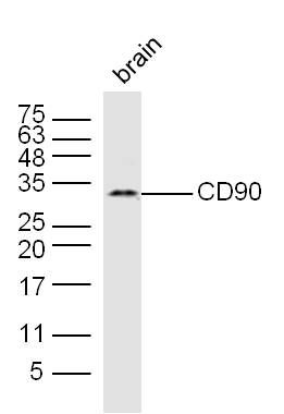 Sample: Sample:Brain (Mouse) Lysate at 40 ug Primary: Anti-CD90 (bs-0778R) at 1/300 dilution Secondary: IRDye800CW Goat Anti-Rabbit IgG at 1/20000 dilution Predicted band size: 12 kD Observed band size: 32 kD 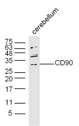 Sample: Sample:Brain (Mouse) Lysate at 40 ug Primary: Anti-CD90 (bs-0778R) at 1/300 dilution Secondary: IRDye800CW Goat Anti-Rabbit IgG at 1/20000 dilution Predicted band size: 12 kD Observed band size: 32 kD Tissue/cell: rat brain tissue; 4% Paraformaldehyde-fixed and paraffin-embedded; Antigen retrieval: citrate buffer ( 0.01M, pH 6.0 ), Boiling bathing for 15min; Block endogenous peroxidase by 3% Hydrogen peroxide for 30min; Blocking buffer (normal goat serum,C-0005) at 37℃ for 20 min; Incubation: Anti-Thy-1/CD90/ Thy1.1 Polyclonal Antibody, Unconjugated(bs-0778R) 1:200, overnight at 4°C, followed by conjugation to the secondary antibody(SP-0023) and DAB(C-0010) staining 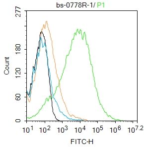 Blank control:Mouse brain. Blank control:Mouse brain.Primary Antibody (green line): Rabbit Anti-CD90 antibody (bs-0778R) Dilution: 1μg /10^6 cells; Isotype Control Antibody (orange line): Rabbit IgG . Secondary Antibody : Goat anti-rabbit IgG-AF488 Dilution: 1μg /test. Protocol The cells were incubated in 5%BSA to block non-specific protein-protein interactions for 30 min at room temperature .Cells stained with Primary Antibody for 30 min at room temperature. The secondary antibody used for 40 min at room temperature. Acquisition of 20,000 events was performed. 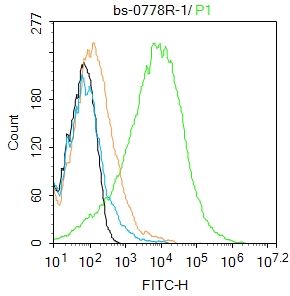 Blank control:Mouse brain. Blank control:Mouse brain.Primary Antibody (green line): Rabbit Anti-CD90 antibody (bs-0778R) Dilution: 1μg /10^6 cells; Isotype Control Antibody (orange line): Rabbit IgG . Secondary Antibody : Goat anti-rabbit IgG-AF488 Dilution: 1μg /test. Protocol The cells were incubated in 5%BSA to block non-specific protein-protein interactions for 30 min at room temperature .Cells stained with Primary Antibody for 30 min at room temperature. The secondary antibody used for 40 min at room temperature. Acquisition of 20,000 events was performed. 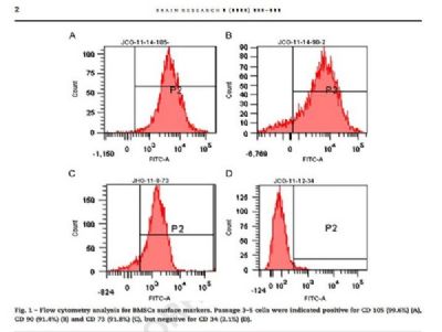 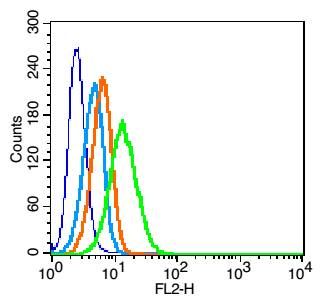 Blank control: U937(blue). Blank control: U937(blue).Primary Antibody: Rabbit Anti-CD90 antibody(bs-0778R), Dilution: 1μg in 100 μL 1X PBS containing 0.5% BSA; Isotype Control Antibody: Rabbit IgG (orange) ,used under the same conditions. Secondary Antibody: Goat anti-rabbit IgG-PE(white blue), Dilution: 1:200 in 1 X PBS containing 0.5% BSA. Protocol The cells were fixed with 2% paraformaldehyde (10 min).Primary antibody (bs-0778R, 1μg /1x10^6 cells) were incubated for 30 min on the ice, followed by 1 X PBS containing 0.5% BSA + 10% goat serum (15 min) to block non-specific protein-protein interactions. Then the Goat Anti-rabbit IgG/PE antibody was added into the blocking buffer mentioned above to react with the primary antibody at 1/200 dilution for 30 min on ice. Acquisition of 20,000 events was performed. 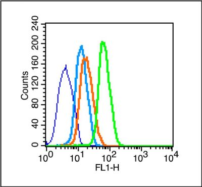 Blank control (blue line): U251 (blue). Blank control (blue line): U251 (blue).Primary Antibody (green line): Rabbit Anti- CD90 antibody (bs-0778R) Dilution: 1μg /10^6 cells; Isotype Control Antibody (orange line): Rabbit IgG . Secondary Antibody (white blue line): Goat anti-rabbit IgG-FITC Dilution: 1μg /test. Protocol The cells were fixed with 70% ice-cold methanol overnight at 4℃. Cells stained with Primary Antibody for 30 min at room temperature. The cells were then incubated in 1 X PBS/2%BSA/10% goat serum to block non-specific protein-protein interactions followed by the antibody for 15 min at room temperature. The secondary antibody used for 40 min at room temperature. Acquisition of 20,000 events was performed. |