上海细胞库
人源细胞系| 稳转细胞系| 基因敲除株| 基因点突变细胞株| 基因过表达细胞株| 重组细胞系| 猪的细胞系| 马细胞系| 兔的细胞系| 犬的细胞系| 山羊的细胞系| 鱼的细胞系| 猴的细胞系| 仓鼠的细胞系| 狗的细胞系| 牛的细胞| 大鼠细胞系| 小鼠细胞系| 其他细胞系|

| 规格 | 价格 | 库存 |
|---|
| 英文名称 | NALP3/CIAS1 |
| 中文名称 | 细胞凋亡诱导蛋白NALP3抗体 |
| 别 名 | LRR and PYD domains-containing protein 3; AGTAVPRL; AII/AVP antibody Angiotensin/vasopressin receptor AII/AVP like; Angiotensin/vasopressin receptor AII/AVP-like; C1orf7; Caterpiller protein 1.1; CIAS 1; CIAS1; CLR1.1; Cold autoinflammatory syndrome 1; Cold autoinflammatory syndrome 1 protein; Cryopyrin; Familial cold autoinflammatory syndrome; FCAS; FCU; Muckle-Wells syndrome; MWS; NACHT; NACHT LRR and PYD containing protein 3; NALP 3; NALP3; NALP3_HUMAN; NLRP3; PYPAF 1; PYPAF1 antibody PYRIN containing APAF1 like protein 1; PYRIN-containing APAF1-like protein 1. |
| 研究领域 | 肿瘤 心血管 细胞生物 免疫学 信号转导 细胞凋亡 转录调节因子 |
| 抗体来源 | Rabbit |
| 克隆类型 | Polyclonal |
| 交叉反应 | Human, Mouse, Rat, Dog, |
| 产品应用 | WB=1:500-2000 ELISA=1:500-1000 IHC-P=1:100-500 IHC-F=1:100-500 ICC=1:100-500 IF=1:200-800 (石蜡切片需做抗原修复) not yet tested in other applications. optimal dilutions/concentrations should be determined by the end user. |
| 分 子 量 | 114kDa |
| 细胞定位 | 细胞核 细胞浆 分泌型蛋白 |
| 性 状 | Liquid |
| 浓 度 | 1mg/ml |
| 免 疫 原 | KLH conjugated synthetic peptide derived from human Cryopyrin:15-120/1036 |
| 亚 型 | IgG |
| 纯化方法 | affinity purified by Protein A |
| 储 存 液 | 0.01M TBS(pH7.4) with 1% BSA, 0.03% Proclin300 and 50% Glycerol. |
| 保存条件 | Shipped at 4℃. Store at -20 °C for one year. Avoid repeated freeze/thaw cycles. |
| PubMed | PubMed |
| 产品介绍 | May function as an inducer of apoptosis. Interacts selectively with ASC and this complex may function as an upstream activator of NF-kappa-B signaling. Inhibits TNF-alpha induced activation and nuclear translocation of RELA/NF-KB p65. Also inhibits transcriptional activity of RELA. Activates caspase-1 in response to a number of triggers including bacterial or viral infection which leads to processing and release of IL1B and IL18. Function: May function as an inducer of apoptosis. Interacts selectively with ASC and this complex may function as an upstream activator of NF-kappa-B signaling. Inhibits TNF-alpha induced activation and nuclear translocation of RELA/NF-KB p65. Also inhibits transcriptional activity of RELA. Activates caspase-1 in response to a number of triggers including bacterial or viral infection which leads to processing and release of IL1B and IL18. Subcellular Location: Cytoplasm. Tissue Specificity: Expressed in blood leukocytes. Strongly expressed in polymorphonuclear cells and osteoblasts. Undetectable or expressed at a lower magnitude in B- and T-lymphoblasts, respectively. High level of expression detected in chondrocytes. Detected in non-keratinizing epithelia of oropharynx, esophagus and ectocervix and in the urothelial layer of the bladder. DISEASE: Defects in NLRP3 are the cause of familial cold autoinflammatory syndrome type 1 (FCAS1) [MIM:120100]; also known as familial cold urticaria. FCAS are rare autosomal dominant systemic inflammatory diseases characterized by episodes of rash, arthralgia, fever and conjunctivitis after generalized exposure to cold. Defects in NLRP3 are a cause of Muckle-Wells syndrome (MWS) [MIM:191900]; also known as urticaria-deafness-amyloidosis syndrome. MWS is a hereditary periodic fever syndrome characterized by fever, chronic recurrent urticaria, arthralgias, progressive sensorineural deafness, and reactive renal amyloidosis. The disease may be severe if generalized amyloidosis occurs. Defects in NLRP3 are the cause of chronic infantile neurologic cutaneous and articular syndrome (CINCA) [MIM:607115]; also known as neonatal onset multisystem inflammatory disease (NOMID). CINCA is a rare congenital inflammatory disorder characterized by a triad of neonatal onset of cutaneous symptoms, chronic meningitis and joint manifestations with recurrent fever and inflammation. Similarity: Belongs to the NLRP family. Contains 1 DAPIN domain. Contains 9 LRR (leucine-rich) repeats. Contains 1 NACHT domain. SWISS: Q96P20 Gene ID: 114548 Database links:
Entrez Gene: 114548 Human Entrez Gene: 216799 Mouse Entrez Gene: 287362 Rat Omim: 606416 Human SwissProt: Q96P20 Human SwissProt: Q8R4B8 Mouse Unigene: 159483 Human Unigene: 54174 Mouse
Important Note: This product as supplied is intended for research use only, not for use in human, therapeutic or diagnostic applications. |
| 产品图片 | 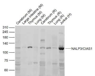 Sample: Sample:Lane 1: Cerebrum (Mouse) Lysate at 40 ug Lane 2: Large intestine (Mouse) Lysate at 40 ug Lane 3: Thymus (Mouse) Lysate at 40 ug Lane 4: Lung (Mouse) Lysate at 40 ug Lane 5: Cerebrum (Rat) Lysate at 40 ug Lane 6: Thymus (Rat) Lysate at 40 ug Lane 7: Lung (Rat) Lysate at 40 ug Lane 8: Jurkat (Human) cell Lysate at 30 ug Primary: Anti-NALP3/CIAS1 (bs-10021R) at 1/1000 dilution Secondary: IRDye800CW Goat Anti-Rabbit IgG at 1/20000 dilution Predicted band size: 118 kD Observed band size: 118 kD 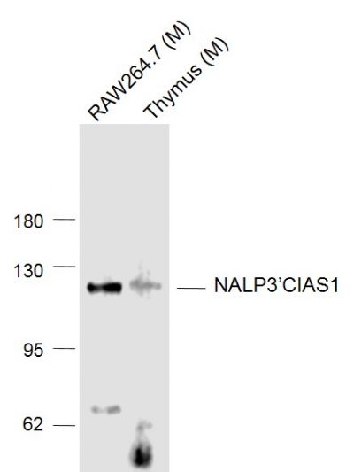 Sample: Sample:Lane 1: RAW264.7 (Mouse) Cell Lysate at 30 ug Lane 2: Thymus (Mouse) Lysate at 40 ug Primary: Anti- NALP3’CIAS1 (bs-10021R) at 1/1000 dilution Secondary: IRDye800CW Goat Anti-Rabbit IgG at 1/20000 dilution Predicted band size: 118 kD Observed band size: 118 kD 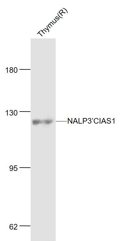 Sample: Sample:Thymus (Rat) Lysate at 40 ug Primary: Anti-NALP3’CIAS1 (bs-10021R) at 1/1000 dilution Secondary: IRDye800CW Goat Anti-Rabbit IgG at 1/20000 dilution Predicted band size: 118 kD Observed band size: 120 kD 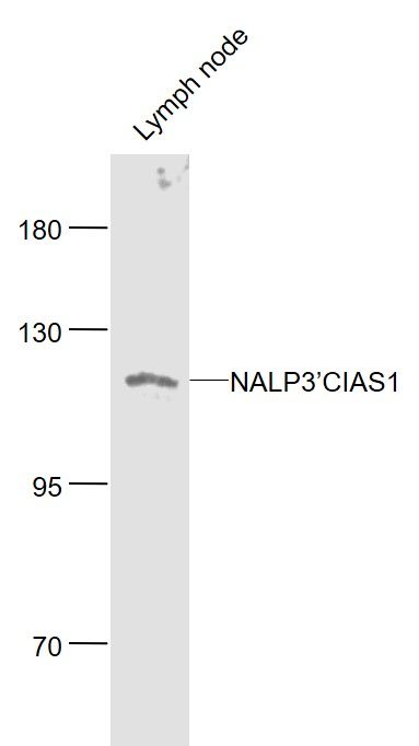 Sample: Sample:Lymph node (Mouse) Lysate at 40 ug Primary: Anti- NALP3’CIAS1 (bs-10021R) at 1/1000 dilution Secondary: IRDye800CW Goat Anti-Rabbit IgG at 1/20000 dilution Predicted band size: 114 kD Observed band size: 114 kD 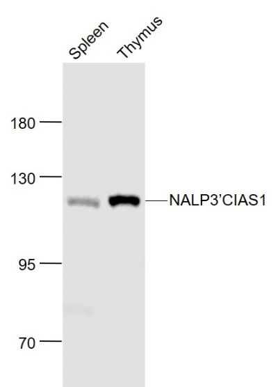 Sample: Sample:Spleen (Mouse) Lysate at 40 ug Thymus (Mouse) Lysate at 40 ug Primary: Anti- NALP3’CIAS1 (bs-10021R) at 1/1000 dilution Secondary: IRDye800CW Goat Anti-Rabbit IgG at 1/20000 dilution Predicted band size: 118 kD Observed band size: 118 kD 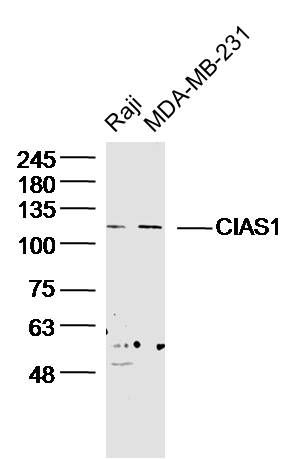 Sample: Sample:Raji Cell (Human) Lysate at 30 ug MDA-MB-231 Cell (Human) Lysate at 30 ug Primary: Anti- CIAS1 (bs-10021R) at 1/300 dilution Secondary: IRDye800CW Goat Anti-Rabbit IgG at 1/20000 dilution Predicted band size: 114kD Observed band size: 114kD 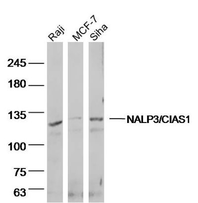 Sample: Sample:Raji (human)cell Lysate at 40 ug MCF-7 (human)cell Lysate at 40 ug siha (human)cell Lysate at 40 ug Primary: Anti- NALP3'CIAS1 (bs-10021R)at 1/300 dilution Secondary: IRDye800CW Goat Anti-Rabbit IgG at 1/20000 dilution Predicted band size: 114kD Observed band size: 114 kD 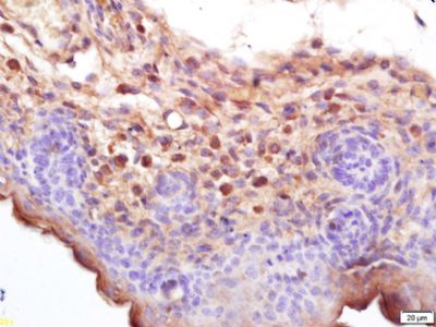 Tissue/cell: mouse embryo tissue; 4% Paraformaldehyde-fixed and paraffin-embedded; Tissue/cell: mouse embryo tissue; 4% Paraformaldehyde-fixed and paraffin-embedded;Antigen retrieval: citrate buffer ( 0.01M, pH 6.0 ), Boiling bathing for 15min; Block endogenous peroxidase by 3% Hydrogen peroxide for 30min; Blocking buffer (normal goat serum,C-0005) at 37℃ for 20 min; Incubation: Anti-Cryopyrin/CIAS1/NALP3 Polyclonal Antibody, Unconjugated(bs-10021R) 1:200, overnight at 4°C, followed by conjugation to the secondary antibody(SP-0023) and DAB(C-0010) staining 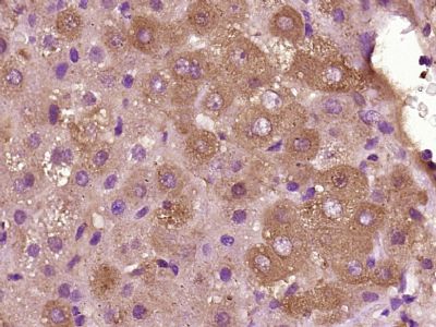 Paraformaldehyde-fixed, paraffin embedded (Human liver cancer); Antigen retrieval by boiling in sodium citrate buffer (pH6.0) for 15min; Block endogenous peroxidase by 3% hydrogen peroxide for 20 minutes; Blocking buffer (normal goat serum) at 37°C for 30min; Antibody incubation with (NALP3/CIAS1) Polyclonal Antibody, Unconjugated (bs-10021R) at 1:400 overnight at 4°C, followed by operating according to SP Kit(Rabbit) (sp-0023) instructionsand DAB staining. Paraformaldehyde-fixed, paraffin embedded (Human liver cancer); Antigen retrieval by boiling in sodium citrate buffer (pH6.0) for 15min; Block endogenous peroxidase by 3% hydrogen peroxide for 20 minutes; Blocking buffer (normal goat serum) at 37°C for 30min; Antibody incubation with (NALP3/CIAS1) Polyclonal Antibody, Unconjugated (bs-10021R) at 1:400 overnight at 4°C, followed by operating according to SP Kit(Rabbit) (sp-0023) instructionsand DAB staining.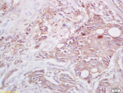 Tissue/cell: human lung carcinoma; 4% Paraformaldehyde-fixed and paraffin-embedded; Tissue/cell: human lung carcinoma; 4% Paraformaldehyde-fixed and paraffin-embedded;Antigen retrieval: citrate buffer ( 0.01M, pH 6.0 ), Boiling bathing for 15min; Block endogenous peroxidase by 3% Hydrogen peroxide for 30min; Blocking buffer (normal goat serum,C-0005) at 37℃ for 20 min; Incubation: Anti-Cryopyrin/CIAS1/NALP3 Polyclonal Antibody, Unconjugated(bs-10021R) 1:200, overnight at 4°C, followed by conjugation to the secondary antibody(SP-0023) and DAB(C-0010) staining 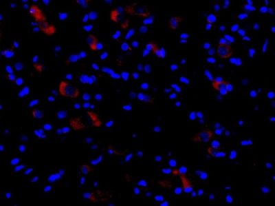 This image was generously provided by Adib Zendedel, PhD from RWTH Aachen University. Formalin-fixed and paraffin embedded rat spinal cord tissue labeled with Rabbit Anti-Cryopyrin Polyclonal Antibody, Unconjugated (bs-10021R) at 1:300 followed by conjugation to a secondary antibody This image was generously provided by Adib Zendedel, PhD from RWTH Aachen University. Formalin-fixed and paraffin embedded rat spinal cord tissue labeled with Rabbit Anti-Cryopyrin Polyclonal Antibody, Unconjugated (bs-10021R) at 1:300 followed by conjugation to a secondary antibody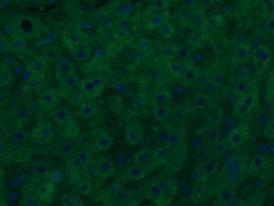 Paraformaldehyde-fixed, paraffin embedded (Human liver cancer); Antigen retrieval by boiling in sodium citrate buffer (pH6.0) for 15min; Block endogenous peroxidase by 3% hydrogen peroxide for 20 minutes; Blocking buffer (normal goat serum) at 37°C for 30min; Antibody incubation with (NALP3/CIAS1) Polyclonal Antibody, Unconjugated (bs-10021R) at 1:400 overnight at 4°C, followed by a conjugated Goat Anti-Rabbit IgG antibody (bs-0295G-AF488) for 90 minutes, and DAPI for nuclei staining. Paraformaldehyde-fixed, paraffin embedded (Human liver cancer); Antigen retrieval by boiling in sodium citrate buffer (pH6.0) for 15min; Block endogenous peroxidase by 3% hydrogen peroxide for 20 minutes; Blocking buffer (normal goat serum) at 37°C for 30min; Antibody incubation with (NALP3/CIAS1) Polyclonal Antibody, Unconjugated (bs-10021R) at 1:400 overnight at 4°C, followed by a conjugated Goat Anti-Rabbit IgG antibody (bs-0295G-AF488) for 90 minutes, and DAPI for nuclei staining. |